
The Inflammation Marker That Can Reveal Trouble Before Symptoms Begin
There is a strange moment in preventive medicine when someone feels healthy, yet the blood suggests another story. Cholesterol may appear acceptable, glucose may remain inside the laboratory range, and blood pressure may seem unremarkable. Nothing hurts, no obvious symptoms appear, and no major diagnosis exists. Then one small laboratory value stands apart: hs-CRP, or high-sensitivity C-reactive protein. That single marker can reveal a level of inflammatory activity that traditional testing may fail to capture.
The most important question is not whether hs-CRP is merely elevated. A better question asks why the body continues producing enough inflammatory signaling to keep that marker high. Inflammation is not inherently harmful because the immune system uses it for healing. Problems begin when the inflammatory response refuses to fully resolve. Instead of protecting damaged tissue temporarily, the immune system can remain activated for weeks, months, or years.
That prolonged activity changes the body’s internal environment. Blood vessels can become more reactive, insulin signaling can weaken, and oxidative stress can increase. Visceral fat may release more inflammatory chemicals, while the liver responds by producing more acute-phase proteins. Over time, that chronic low-grade inflammatory state can contribute to cardiovascular disease and metabolic dysfunction. hs-CRP offers a simple window into that deeper process.
What hs-CRP Actually Measures
C-reactive protein belongs to a group known as acute-phase proteins. The liver increases production when inflammatory signals indicate injury, infection, immune activation, or metabolic stress. Interleukin-6, often called IL-6, plays an important role in stimulating this liver response. Other cytokines also contribute, creating a communication network between stressed tissues, immune cells, and the liver.
A standard CRP test detects larger inflammatory responses, including those associated with infection or major tissue injury. High-sensitivity CRP measures much smaller concentrations with far greater precision. That increased sensitivity makes hs-CRP useful for studying chronic low-grade inflammation, especially in cardiovascular risk assessment. Someone can therefore have an elevated hs-CRP without feeling acutely ill.
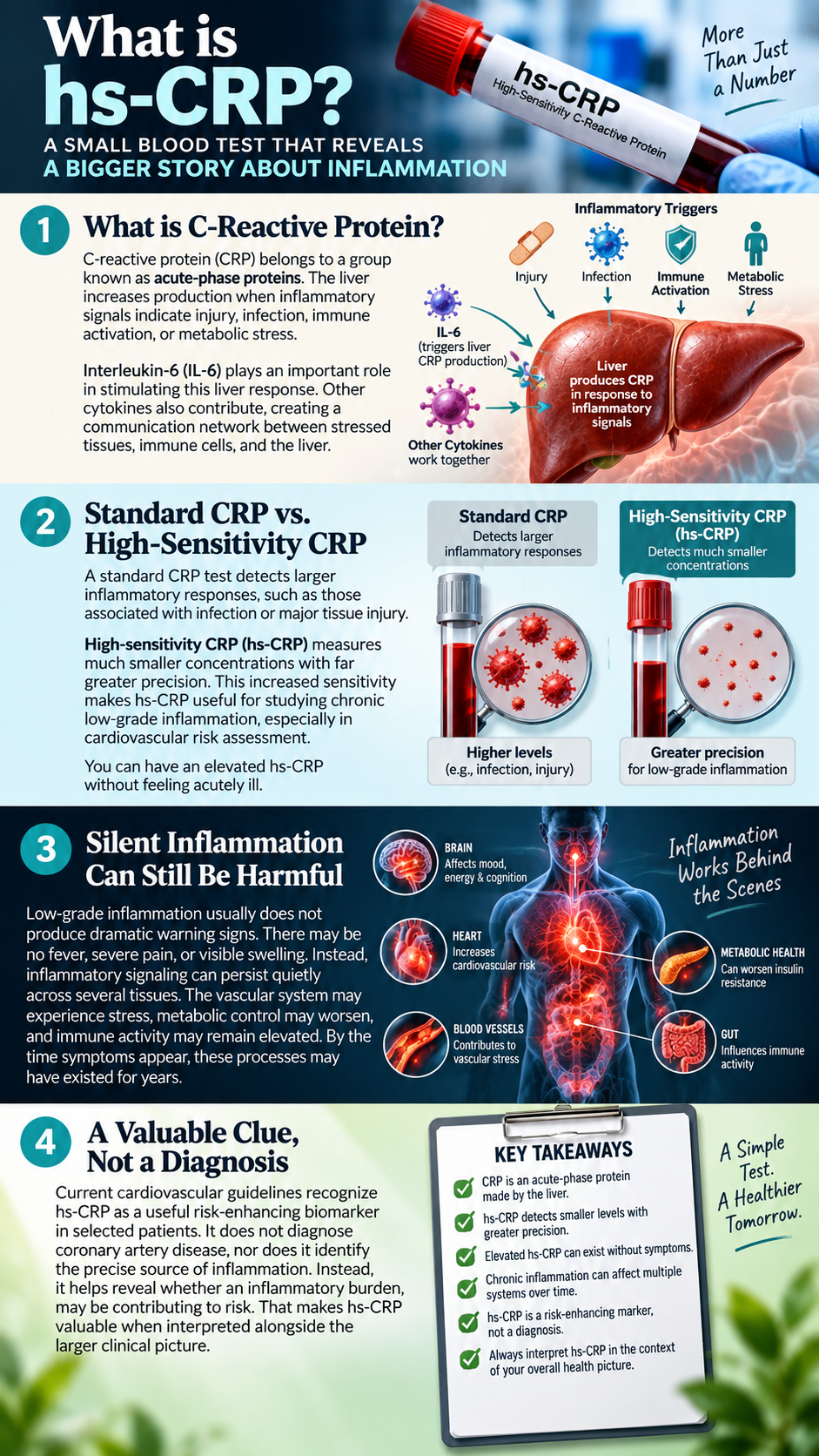
This distinction matters because low-grade inflammation usually does not produce dramatic warning signs. There may be no fever, severe pain, or visible swelling. Instead, inflammatory signaling can persist quietly across several tissues. The vascular system may experience stress, metabolic control may worsen, and immune activity may remain elevated. By the time symptoms appear, these processes may have existed for years.
Current cardiovascular guidelines recognize hs-CRP as a useful risk-enhancing biomarker in selected patients. It does not diagnose coronary artery disease, nor does it identify the precise source of inflammation. Instead, it helps reveal whether an inflammatory burden may be contributing to risk. That makes hs-CRP valuable when interpreted alongside the larger clinical picture.
Understanding the hs-CRP Blood Test
Traditional hs-CRP interpretation divides results into broad cardiovascular risk categories. Levels below 1.0 mg/L have historically represented lower relative inflammatory risk. Measurements between 1.0 and 3.0 mg/L have commonly represented intermediate relative risk. Results above 3.0 mg/L have generally represented higher relative cardiovascular risk.
These ranges should never become rigid labels because hs-CRP reflects more than cardiovascular biology. Infection, injury, intense exercise, dental procedures, inflammatory disorders, and other stressors can temporarily raise CRP. A value that appears concerning during influenza may look completely different several weeks later. Context therefore matters as much as the number itself.
A markedly elevated result requires even greater caution. Levels around 10 mg/L or higher often suggest an acute inflammatory process rather than chronic cardiovascular inflammation alone. In those situations, repeating the test after recovery can provide more meaningful information. One isolated value should rarely become the entire basis for cardiovascular conclusions.
Patterns provide far more insight than single numbers. Elevated hs-CRP alongside high fasting insulin, increasing waist circumference, elevated triglycerides, and worsening blood pressure tells one story. The same hs-CRP after a recent infection tells another. Laboratory medicine becomes far more useful when those numbers form a physiological pattern.
The Real Problem Is Not the CRP Molecule
One of the most common mistakes is treating CRP as though the protein itself represents the disease. It does not. CRP mainly serves as a signal that inflammatory biology is active somewhere in the body. The deeper problem is the environment that caused the liver to increase CRP production.
Imagine a smoke detector sounding in the middle of the night. Removing the batteries might silence the alarm, but it would not extinguish the fire. Treating hs-CRP as the primary problem follows the same faulty logic. The important work involves locating the source of the inflammatory signal.
That source can vary dramatically between people. Visceral fat may be driving inflammatory cytokines in one person. Another may have severe periodontal disease or poorly controlled insulin resistance. Someone else may suffer from chronic sleep disruption, smoking exposure, inflammatory disease, infection, or several factors at once.
A useful investigation therefore works backward from the marker. Rather than asking only how to lower hs-CRP, ask what keeps stimulating inflammatory pathways. Which tissues are stressed, and where is immune activation coming from? What metabolic dysfunction is present, and what daily behaviors may be sustaining it?
Those questions move the conversation away from disease labeling and toward physiology. They also explain why no single supplement can universally fix an elevated hs-CRP. Different biological causes require different interventions. The marker helps reveal the smoke, while a deeper investigation finds the fire.
Visceral Fat: A Hidden Source of Inflammatory Signaling
Most people still imagine body fat as inactive storage. Visceral fat behaves very differently because it surrounds internal organs and communicates with the immune system. This tissue releases hormones, fatty acids, and inflammatory signaling molecules. For that reason, adipose tissue functions as both an endocrine and immunological organ.
Problems develop when fat cells continually absorb more energy than they can safely manage. Adipocytes enlarge, cellular stress increases, and local oxygen delivery can become inadequate. Some cells become dysfunctional, while others eventually die. The immune system then sends macrophages into the tissue to clean up damaged cells.
Those macrophages may surround injured adipocytes and create structures called crown-like formations. Within that environment, inflammatory mediators such as IL-6 and tumor necrosis factor-alpha can increase. A process that begins as tissue repair may eventually become chronic metabolic inflammation. That persistent signaling can extend far beyond the fat tissue itself.
Visceral fat therefore creates more than a cosmetic problem. It can interfere with insulin signaling, influence liver function, and alter lipid metabolism. Increased fatty acid release can place additional stress on the liver and skeletal muscle. Systemic inflammatory markers, including hs-CRP, may rise as these processes continue.
This connection helps explain why central obesity often appears alongside insulin resistance and elevated hs-CRP. The liver is not randomly producing inflammatory proteins. It may be responding to a constant stream of biochemical stress signals. Those signals often originate from metabolically dysfunctional tissues.
Insulin Resistance Can Intensify Inflammation
Blood sugar dysfunction often begins long before diabetes appears on a laboratory report. The pancreas can produce increasingly large amounts of insulin to keep glucose inside the normal range. Fasting glucose may therefore look acceptable while metabolic control quietly deteriorates. That compensated phase can last for years.
As insulin resistance advances, the pancreas continues increasing insulin output. Higher insulin can promote fat storage and reduce access to stored fat. Visceral adipose tissue may expand, while fat can also accumulate inside the liver. Triglycerides frequently rise, and HDL may fall as the metabolic environment changes.
Inflammatory signaling and insulin resistance can then reinforce each other. Certain cytokines interfere with normal insulin pathways inside cells. Poor insulin sensitivity creates additional metabolic stress, which further stimulates inflammatory signaling. Dysfunctional fat tissue contributes even more cytokines to that cycle.
This biological feedback loop helps explain the close relationship between metabolic syndrome and chronic inflammation. The problem involves much more than high blood sugar. Insulin resistance, visceral adiposity, abnormal lipids, vascular stress, and inflammation often develop together. hs-CRP may provide one important clue that this larger network is active.
What Chronic Inflammation Does to the Arteries
Arteries are living biological structures, not passive pipes. Their inner lining contains endothelial cells that regulate blood flow, vascular tone, clotting, and immune interactions. These cells constantly respond to the metabolic environment passing across them. Chronic inflammatory signals can disturb that delicate balance.
When vascular tissue becomes stressed, immune-cell recruitment can increase. Lipoproteins entering vulnerable arterial tissue may undergo chemical modification. Macrophages then enter the arterial wall and participate in lipid handling. Over time, atherosclerotic plaque can develop through the interaction of lipid exposure and inflammation.
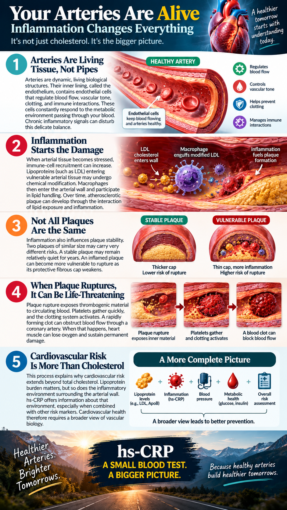
Inflammation also influences plaque stability. Two plaques of similar size may carry very different risks. A stable plaque may remain relatively quiet for years. An inflamed plaque can become more vulnerable to rupture as its protective fibrous cap weakens.
Plaque rupture exposes thrombogenic material to circulating blood. Platelets gather quickly, and the clotting system activates. A rapidly forming clot can obstruct blood flow through a coronary artery. When that happens, heart muscle can lose oxygen and sustain permanent damage.
This process explains why cardiovascular risk extends beyond total cholesterol. Lipoprotein burden matters, but so does the inflammatory environment surrounding the arterial wall. hs-CRP offers information about that environment, especially when combined with other risk markers. Cardiovascular health therefore requires a broader view of vascular biology.
Your Mouth May Be Contributing to Systemic Inflammation
The mouth is often excluded from conversations about cardiovascular health. That separation makes little biological sense because gum tissue contains blood vessels, bacteria, immune cells, and inflammatory pathways. Chronic periodontal disease can therefore create an ongoing inflammatory burden. The effects may extend far beyond the teeth and gums.
Periodontitis develops when chronic inflammation damages the tissues supporting teeth. Deep periodontal pockets can harbor bacterial communities that repeatedly stimulate immune activity. Bleeding gums provide a visible clue that this tissue remains inflamed. Lack of pain does not mean the process is harmless.
Research has repeatedly linked periodontal disease with elevated CRP and hs-CRP. Treatment studies have also shown reductions in systemic inflammatory markers after periodontal therapy. That does not prove gum disease causes every cardiovascular event. It does show that oral inflammation can contribute to measurable systemic inflammation.
This connection offers an important lesson about root-cause investigation. A cardiovascular inflammation marker may partly reflect pathology inside the mouth. Treating inflamed periodontal tissue can reduce one source of inflammatory signaling. That makes oral health an important part of any serious inflammation assessment.
Poor Sleep Can Keep Inflammatory Pathways Active
Sleep is one of the most underestimated regulators of inflammation. The sleeping body is not inactive because numerous repair and regulatory processes occur overnight. Hormonal rhythms shift, immune signaling changes, and autonomic activity moves toward recovery. Glucose regulation and tissue repair also depend on adequate sleep.
Repeated sleep restriction disrupts many of those processes. Experimental research shows that short sleep can increase inflammatory markers such as IL-6 and CRP. The effect becomes particularly noticeable after several consecutive nights of insufficient sleep. Chronic sleep disruption may therefore create a persistent inflammatory burden.
Obstructive sleep apnea deserves even greater attention. Repeated airway obstruction can lower oxygen levels and fragment normal sleep architecture. The brain and cardiovascular system experience repeated episodes of physiological stress. Someone may spend eight hours in bed yet receive little restorative sleep.
Symptoms such as loud snoring, gasping, morning headaches, and severe daytime sleepiness deserve investigation. Ignoring these signs can leave a major source of inflammatory stress untreated. When hs-CRP remains elevated, sleep quality should always be considered. Better sleep can support healthier immune and metabolic regulation.
Stop Chasing the Marker and Start Investigating the Terrain
Imagine finding water across your kitchen floor every morning. Measuring the depth would provide information, but it would not repair the leaking pipe. An elevated hs-CRP deserves the same kind of thinking. The laboratory value matters, yet the underlying cause matters far more.
A strong investigation begins by identifying likely sources of chronic inflammatory signaling. Visceral fat, insulin resistance, periodontal disease, poor sleep, smoking, chronic infection, and inflammatory disorders deserve consideration. Dietary patterns and physical inactivity may also contribute. In many cases, several drivers operate at the same time.
This complexity explains why one intervention rarely solves everything. Someone may improve their diet while ignoring severe periodontal disease. Another person may exercise daily while sleeping five hours each night. A third may lose weight while continuing to smoke.
Meaningful improvement requires attention to the entire biological terrain. The objective is not simply lowering hs-CRP on the next laboratory test. The real goal is reducing the inflammatory burden that caused the elevation. When that environment changes, the marker often follows.
Build Meals That Support Metabolic Stability
Food represents one of the most controllable influences on metabolic health. That does not mean everyone needs the same diet. Instead, meals should support stable glucose, healthy insulin signaling, appropriate body composition, and adequate nutrient intake. Those goals matter more than following a fashionable diet label.
Refined carbohydrates and sweetened drinks can create repeated glucose surges in susceptible people. The pancreas responds by releasing insulin again and again. Constant snacking can extend that insulin exposure throughout the day. Over time, this pattern may contribute to worsening insulin resistance.
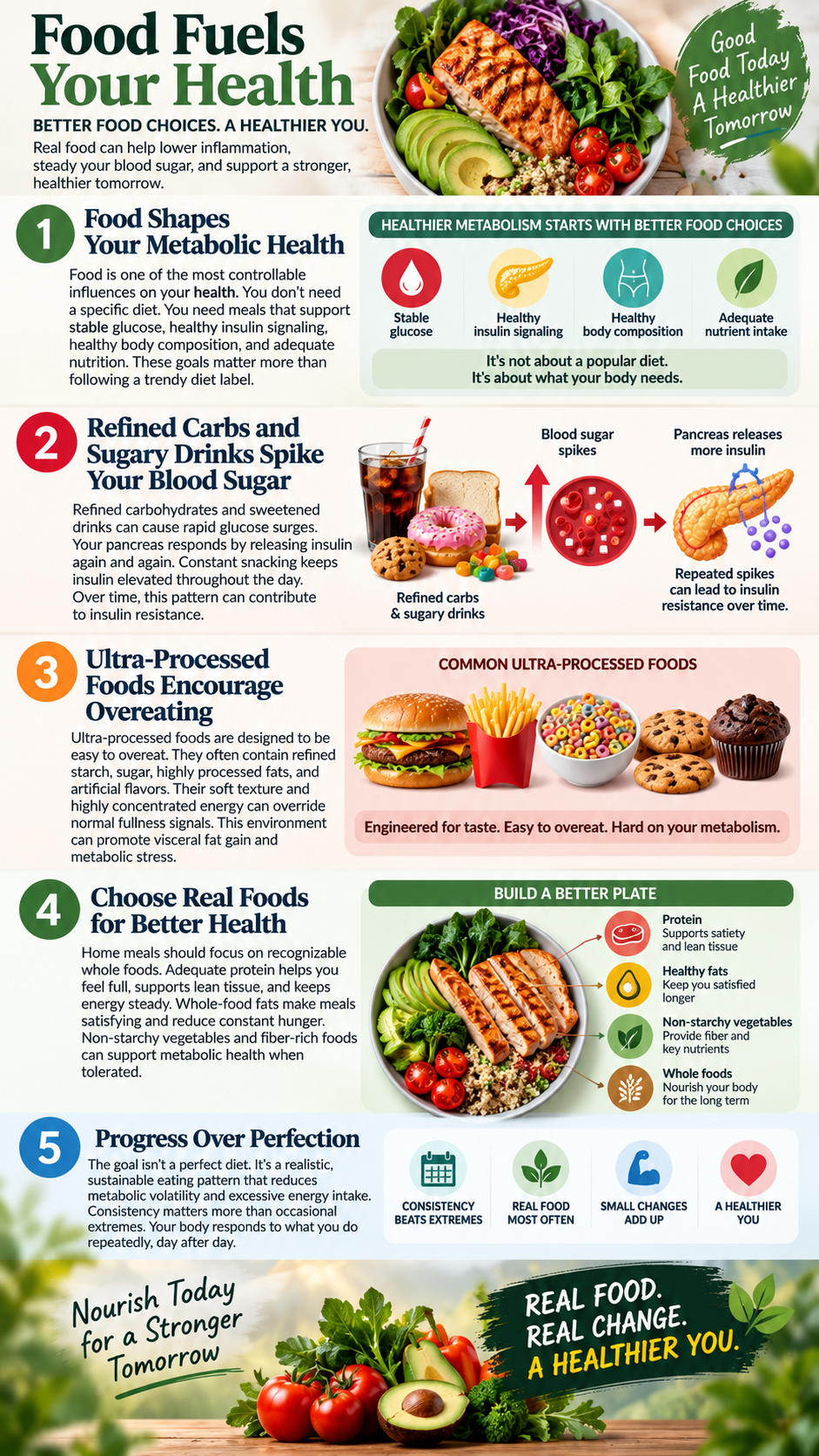
Ultra-processed foods create another problem because they encourage overeating. Many combine refined starch, sugar, highly processed fats, and engineered flavors. Their soft texture and concentrated energy make excessive consumption remarkably easy. That environment can promote visceral fat gain and metabolic stress.
Home meals should focus primarily on recognizable whole foods. Adequate protein can support satiety, lean tissue, and stable energy. Whole-food fats can make meals satisfying and reduce constant hunger. Non-starchy vegetables and other fiber-rich foods can support metabolic health when tolerated.
The goal should never be perfection. Instead, create a food pattern that reduces metabolic volatility and excessive energy intake. Consistency matters far more than occasional dietary extremes. The body responds to what happens repeatedly.
Give Insulin Time to Fall Between Meals
Modern eating patterns often begin shortly after waking and continue until bedtime. Breakfast leads to snacks, lunch creates another snack, and dinner ends with dessert. The digestive and endocrine systems receive very little pause. That constant feeding environment keeps insulin elevated more frequently.
Many adults can benefit from eliminating unnecessary grazing between meals. Several hours between meals can allow insulin levels to decline. That change may also improve appetite awareness and reduce automatic eating. People often discover that many snacks came from habit rather than hunger.
Time-restricted eating can provide another useful strategy for selected individuals. A consistent eating window may reduce late-night food intake and improve metabolic organization. The approach should remain flexible because not everyone responds well to prolonged fasting. Medical conditions and individual needs always matter.
A practical first step involves finishing dinner earlier and avoiding nighttime snacking. That change shortens the eating window without creating an aggressive fasting protocol. Sustainable improvements usually outperform extreme strategies. The objective is better metabolic regulation, not endurance testing.
Reduce Visceral Fat Without Obsessing Over Body Weight
A bathroom scale cannot distinguish muscle from visceral fat. It also cannot separate bone, water, or subcutaneous fat from metabolically harmful fat stores. For that reason, body weight alone provides incomplete information. Waist circumference often offers additional insight into central adiposity.
Reducing visceral fat can lower several sources of inflammatory signaling. However, muscle preservation should remain a priority during fat loss. Skeletal muscle helps clear glucose from the bloodstream and supports insulin sensitivity. Losing large amounts of muscle can undermine long-term metabolic health.
Resistance training deserves an important role because it protects and builds lean tissue. Squats, rows, presses, hinges, and carries can recruit large muscle groups. Beginners can perform modified versions using body weight or resistance bands. Progress should remain steady rather than aggressive.
Walking provides another powerful but underrated tool. A short walk after meals can help muscles use circulating glucose. Longer daily walks also support energy expenditure and cardiovascular fitness. Frequent movement throughout the day can matter as much as structured workouts.
Fat loss works best when habits remain sustainable. Severe calorie restriction often increases hunger and sacrifices muscle. Rebound eating can quickly erase short-term progress. Slow improvements in body composition usually create more durable metabolic changes.
Treat Oral Health as Part of Whole-Body Health
Bleeding gums should never become normal. Persistent bleeding indicates inflamed tissue, even when the gums do not hurt. That inflammation deserves proper evaluation. Ignoring it can allow periodontal disease to progress silently.
Daily oral care remains essential for controlling bacterial buildup. Thorough brushing twice daily helps disrupt plaque around teeth and gums. Cleaning between teeth removes debris from areas toothbrushes rarely reach. Floss or interdental brushes can support this process.
Advanced periodontal disease cannot be corrected through home care alone. Deep pockets, bone loss, and significant gum recession require professional evaluation. Periodontal treatment can reduce local inflammation and bacterial burden. Research also suggests that systemic CRP may decline after successful therapy.
That relationship reinforces an important principle. Oral health belongs inside the larger conversation about systemic inflammation. The mouth is not separate from the cardiovascular system. Chronic gum disease can contribute to the inflammatory load carried throughout the body.
Make Sleep an Anti-Inflammatory Practice
Many people try to correct exhaustion with caffeine instead of improving sleep. That strategy creates temporary alertness without repairing the underlying problem. The immune and metabolic systems still experience the consequences of sleep loss. Eventually, the body collects that debt.
Most adults should aim for seven to nine hours of sleep opportunity. Consistent bedtimes and waking times can strengthen circadian regulation. Morning outdoor light can also support healthy timing signals. Evening darkness helps prepare the brain for sleep.
The bedroom environment matters as well. A cool, dark, and quiet room usually supports deeper sleep. Heavy meals late at night can disturb rest in some people. Alcohol may create initial drowsiness while fragmenting later sleep.
Persistent snoring or gasping deserves careful attention. Severe daytime sleepiness should also raise concern about sleep apnea. Lifestyle improvements cannot fully correct repeated airway obstruction. Proper evaluation can uncover an important inflammatory stressor.
Move Every Day but Respect Recovery
Exercise improves glucose disposal, cardiovascular fitness, body composition, and metabolic resilience. However, exercise works best when paired with adequate recovery. Excessive training without sufficient sleep can temporarily increase inflammatory stress. The body needs time to repair stressed tissues.
Daily movement should begin with walking and frequent activity. Sitting for ten consecutive hours creates a metabolic burden that one evening workout cannot fully erase. Regular movement throughout the day supports glucose control and circulation. Short activity breaks can provide meaningful benefits.
Strength training should also become part of the weekly routine. Building muscle improves glucose handling and supports long-term metabolic health. Aerobic conditioning improves mitochondrial function and cardiovascular capacity. Combining both approaches creates broader benefits than relying on one method.
Testing hs-CRP immediately after unusually hard exercise can also distort interpretation. Strenuous training temporarily increases inflammatory signals as the body repairs muscle tissue. Avoiding intense exercise before testing can provide a cleaner picture. Context always improves laboratory interpretation.
Remove the Obvious Inflammatory Inputs
Cigarette smoke remains one of the clearest avoidable inflammatory exposures. Smoking damages endothelial function and increases oxidative stress. It also accelerates several processes involved in cardiovascular disease. No supplement can neutralize ongoing smoking.
Excessive alcohol can create another significant burden. Frequent heavy drinking can disrupt sleep, liver function, glucose regulation, appetite, and blood pressure. Those changes can increase metabolic and inflammatory stress. Reducing excessive intake can improve several systems at once.
Ultra-processed foods should also become occasional rather than foundational. Repeated overeating can expand visceral fat regardless of whether individual ingredients seem healthy. Total metabolic context matters more than marketing claims. Whole foods make energy regulation easier for many people.
Chronic psychological stress deserves attention for similar reasons. Persistent sympathetic activation can disrupt sleep, appetite, glucose regulation, and blood pressure. Practices such as prayer, meditation, breathing exercises, and outdoor time can support healthier stress physiology. Strong social relationships can provide similar benefits.
Retest hs-CRP and Look for Patterns
A single blood draw captures one moment in time. Health represents a changing trajectory rather than one isolated measurement. Repeat testing can therefore provide valuable information when the first hs-CRP appears unexpectedly elevated. Timing should account for temporary inflammatory events.
Avoid testing during acute illness when cardiovascular risk assessment is the goal. Recent injury, dental procedures, or intense exercise may also elevate CRP temporarily. Once those factors resolve, repeat testing can reveal whether inflammation remains elevated. Persistent elevation deserves deeper investigation.
Interpret the follow-up value alongside other metabolic markers. Fasting insulin, triglycerides, HDL, glucose, A1c, blood pressure, ApoB, and waist circumference can add important context. Liver markers and lipoprotein(a) may also help refine risk. No single test should carry the entire burden of interpretation.
Improvement becomes especially meaningful when several markers change together. Lower hs-CRP combined with better insulin, lower triglycerides, and reduced waist circumference tells a stronger story. Increased fitness and improved sleep add further confirmation. That pattern suggests the internal environment truly changed.
hs-CRP Is an Invitation to Look Deeper
The greatest value of hs-CRP may lie outside the number itself. This simple blood test reminds us that chronic disease rarely begins when symptoms first appear. Biology changes quietly long before many diagnoses become obvious. Early markers create an opportunity to investigate before damage advances.
Inflammation can grow within visceral fat, arterial tissue, diseased gums, and other stressed tissues. Sleep loss can amplify immune signaling, while insulin resistance can deepen metabolic dysfunction. Several pathways may eventually converge. The result is a body operating in a chronically defensive state.
That environment can affect cardiovascular health, glucose regulation, energy production, and tissue recovery. Blood vessels may become less resilient, while metabolic flexibility declines. Visceral fat can continue expanding as inflammatory signaling grows. The process can remain silent until a major diagnosis finally appears.
hs-CRP should therefore inspire curiosity rather than fear. One elevated test does not predict an inevitable heart attack or stroke. Instead, it provides a reason to examine the body’s inflammatory terrain. The smartest response involves identifying probable sources and changing what can be changed.
Improve metabolic health, protect sleep, strengthen muscle, and reduce visceral fat. Address gum disease and stop smoking when those factors apply. Replace ultra-processed foods with meals that support metabolic stability. Then retest and watch for changes across the entire physiological pattern.
A laboratory report should never become the final chapter of someone’s health story. Used intelligently, hs-CRP can become the beginning of a much more useful investigation. The real question is not simply how high the marker appears. The better question asks where the inflammation comes from and how that environment can change.