
THE INFECTION HIDING IN PLAIN SIGHT
There is something deeply unsettling about an infection that can live inside the body for decades without announcing itself. No dramatic fever necessarily marks the moment of infection, and no unmistakable illness always follows. Instead, a microscopic organism can quietly occupy the stomach while life keeps moving forward. Childhood becomes adolescence, careers begin, families grow, and seemingly unrelated symptoms gradually appear. The infection remains hidden beneath digestive complaints that may receive simpler explanations.
That organism is Helicobacter pylori, commonly called H. pylori. It is a spiral-shaped bacterium capable of living inside the stomach’s protective mucus layer. Researchers once believed bacteria could barely survive the stomach’s highly acidic environment, but H. pylori shattered that assumption. Its discovery rewrote our understanding of ulcers, gastritis, and gastric cancer. Today, its significance extends far beyond occasional indigestion or stomach discomfort.
Many infected people never develop obvious symptoms, while others experience bloating, nausea, abdominal burning, excessive belching, reflux-like symptoms, or chronic indigestion. Some develop ulcers, iron deficiency, reduced appetite, or long-standing gastritis. Unfortunately, these complaints often get treated as separate problems instead of being connected to a deeper underlying process. The result can become years of symptom management without identifying the bacterial infection driving inflammation.
The real problem is not simply that bacteria entered the stomach. The greater concern involves what happens when the infection creates years of unresolved gastric inflammation. Chronic inflammation can alter stomach acid production, damage protective tissue, disturb nutrient handling, and change cellular behavior. In susceptible people, these changes may eventually raise the risk of serious gastric disease.
The National Cancer Institute recognizes chronic H. pylori infection as a major cause of persistent stomach inflammation. It also links the organism with peptic ulcers, gastric adenocarcinoma, and gastric MALT lymphoma. This does not mean every infected person develops cancer or ulcers, but it does mean the infection deserves more attention than another antacid prescription.
How H. pylori Found a Home Inside the Human Stomach
For decades, medical textbooks portrayed the stomach as an almost impossible environment for bacterial survival. Hydrochloric acid creates a hostile setting capable of destroying countless organisms entering with food and water. Scientists therefore assumed stomach ulcers resulted mainly from stress, spicy food, excess acid, or emotional tension. That theory dominated medicine until two Australian researchers began questioning what everyone else had accepted.
During the early 1980s, Barry Marshall and Robin Warren identified curved bacteria inside stomach tissue from patients with gastritis. Their work eventually established H. pylori as a major driver of chronic gastritis and peptic ulcer disease. The discovery transformed gastroenterology and ultimately earned them the 2005 Nobel Prize in Physiology or Medicine. Their research also exposed an important lesson about medicine itself: longstanding assumptions can survive for years until someone looks more carefully at the underlying biology.
The organism, however, had already spent thousands of years adapting to human beings. Most infections appear to begin during childhood, particularly in environments where close household contact increases exposure. Transmission can occur through contact with infected saliva, vomit, or fecal material. Household clustering has also been documented, which helps explain why more than one person in a family may carry the organism.
Once H. pylori reaches the stomach, survival becomes its first challenge. The organism overcomes that challenge with remarkable biochemical precision. It does not simply endure stomach acid through brute resistance. Instead, it manipulates its surroundings and moves toward a more protected location near the gastric lining.
The Bacterium That Learned to Survive Stomach Acid
H. pylori produces an enzyme called urease, which helps it survive the stomach’s highly acidic environment. Urease breaks down urea and creates compounds that reduce acidity immediately around the bacterium. This process does not neutralize the whole stomach, but it creates a localized chemical shield. That tiny protective zone gives the organism enough time to survive and migrate deeper into the mucus layer.
Its corkscrew shape and flagella provide another advantage. These structures help H. pylori move through gastric mucus and approach the epithelial cells underneath. Once it reaches that protected environment, it attaches to the stomach lining and becomes more difficult to eliminate. The organism has effectively moved away from the harshest acid exposure and into a niche where survival becomes far more likely.
The immune system quickly recognizes that something foreign has arrived. Immune cells gather around infected tissue and release inflammatory compounds designed to destroy the invader. Yet H. pylori interferes with local immune responses and often prevents complete elimination. The result becomes a chronic biological stalemate in which the immune system continues attacking while the organism continues surviving.
That chronicity changes the entire picture. Instead of a short infection followed by recovery, the stomach can enter a prolonged state of inflammatory stress. Years of immune activation can alter tissue structure, change acid production, and weaken normal protective mechanisms. The longer the infection persists, the greater the opportunity for deeper physiological disruption.
The Real Problem Is the Chronic Inflammatory Environment
Finding H. pylori answers one important question, but it raises another. What has the organism been doing to the stomach during all those years? The answer depends partly on where the infection sits, how severe the inflammation becomes, and how the individual body responds.
The stomach is far more than a storage chamber for food. It produces hydrochloric acid, digestive enzymes, signaling molecules, intrinsic factor, and protective mucus. These systems help begin protein digestion, release nutrients from food, regulate bacterial exposure, and prepare the small intestine for what comes next. When chronic gastritis disrupts those functions, the consequences can spread well beyond the stomach.
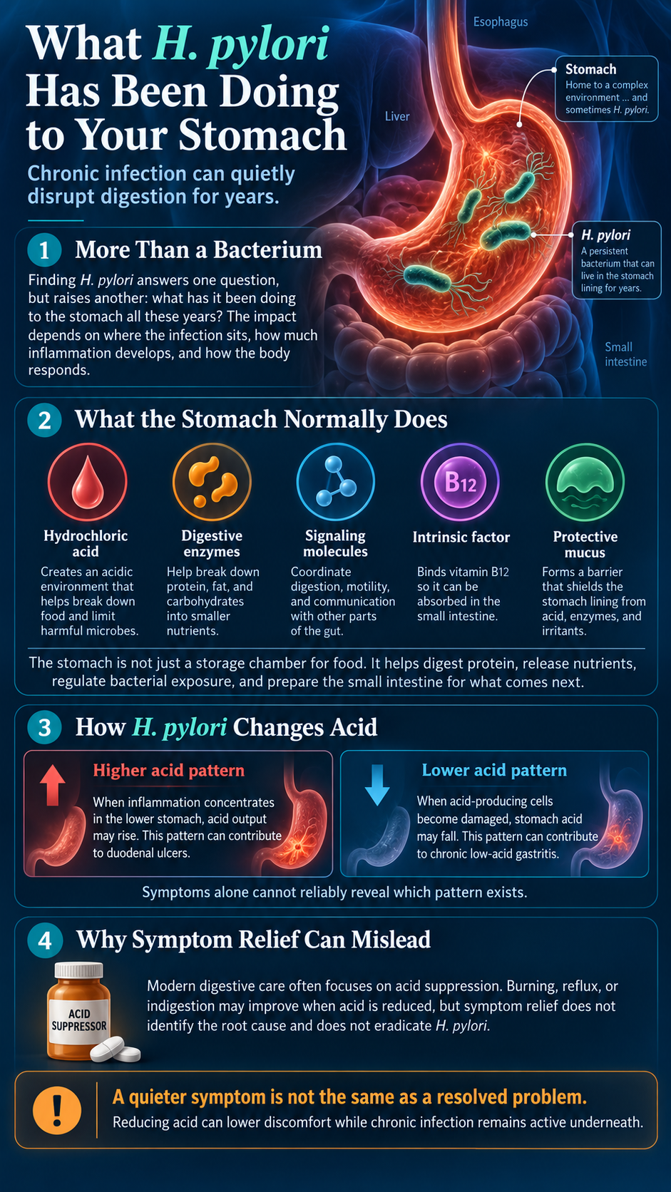
Some patterns of H. pylori infection can increase acid output, especially when inflammation concentrates in the lower stomach. Other patterns eventually damage acid-producing cells and reduce acid secretion. This difference explains why one infected person may develop duodenal ulcers while another develops low-acid gastritis. Symptoms alone cannot reliably reveal which pattern exists.
That distinction matters because modern digestive care often revolves around acid suppression. Someone develops burning, reflux, or indigestion and receives a medication designed to reduce stomach acid. The symptoms may improve because less acid contacts irritated tissue. However, symptom relief does not identify the underlying cause, and it certainly does not eradicate H. pylori.
A quieter symptom is not always the same as a resolved problem. Reducing acid can lower discomfort while chronic infection remains active underneath. That difference matters when someone uses acid-suppressing medication for months or years without ever investigating why the stomach became inflamed in the first place.
When Chronic Gastritis Starts Changing Digestion
Healthy stomach acid performs several essential jobs. It begins protein digestion, activates digestive enzymes, helps liberate nutrients from food, and creates a chemical barrier against swallowed microbes. Gastric acidity also supports iron and vitamin B12 physiology through several mechanisms. When chronic gastritis changes acid production, digestion can become less efficient even when symptoms remain subtle.
Longstanding H. pylori infection can contribute to iron deficiency in susceptible individuals. Several mechanisms may participate, including altered gastric acidity, reduced iron availability, microscopic blood loss, and bacterial competition for iron. Current gastroenterology guidelines recognize unexplained iron-deficiency anemia as an indication for H. pylori evaluation.
Vitamin B12 status can also deserve attention when extensive gastric damage develops. Severe atrophic gastritis may interfere with the stomach environment necessary for normal B12 absorption. Over time, deficiencies may show up as fatigue, weakness, cognitive changes, numbness, or anemia. The original stomach infection may no longer look like a digestive problem at all.
This is one reason H. pylori can remain hidden for so long. The downstream symptoms may appear in other systems while the origin remains gastric. A person may chase fatigue, low iron, poor concentration, or unexplained weakness without anyone asking whether chronic stomach inflammation sits underneath the pattern.
H. pylori and the Ulcer Connection
Few discoveries changed ulcer treatment as dramatically as the identification of H. pylori. Before the bacterial connection became widely accepted, ulcer care focused heavily on stress, diet, and excess acid. Patients received bland diets, antacids, acid suppressors, and sometimes surgery. Many improved temporarily, yet recurrent disease remained common.
The underlying infection helped explain those recurrences. Chronic gastritis weakens the stomach’s protective defenses, disrupts mucus production, and alters inflammatory signaling. In some people, acid exposure then injures already vulnerable tissue. Over time, that injury can deepen enough to create an ulcer within the stomach or duodenum.
Ulcers can cause burning abdominal pain, nausea, nighttime discomfort, early fullness, or pain after meals. Bleeding ulcers may produce dark stools, vomiting of blood, or slowly developing iron-deficiency anemia. Some people experience very little discomfort until bleeding becomes significant. This variation makes symptom severity an unreliable measure of tissue damage.
NSAID medications create another layer of risk because they weaken protective mechanisms within the gastrointestinal lining. Someone carrying H. pylori while frequently using NSAIDs may face overlapping sources of injury. That combination illustrates a broader truth about chronic disease: multiple stressors often converge long before symptoms become severe.
The Silent Progression Toward More Serious Disease
The most serious consequence of chronic H. pylori infection may unfold without dramatic symptoms. Persistent inflammation can gradually transform stomach tissue through a sequence of biological changes. The process often begins with chronic gastritis and may advance toward atrophic gastritis in susceptible individuals.
Atrophic gastritis involves loss of normal gastric glands and declining function of the stomach lining. Intestinal metaplasia can follow, causing stomach cells to take on characteristics resembling intestinal tissue. Additional cellular abnormalities may appear later in a smaller percentage of patients. This progression does not occur in everyone, but it helps explain the connection between chronic infection and gastric cancer.
H. pylori is classified as a human carcinogen because the relationship with gastric cancer is well established. Chronic infection increases the risk of gastric adenocarcinoma and gastric MALT lymphoma. Research also shows that eradicating the infection can reduce gastric cancer risk in several populations. (cancer.gov)
Bacterial strain also matters because some strains carry more aggressive virulence factors. Certain H. pylori strains produce CagA, a protein associated with stronger inflammatory effects and greater cancer risk. Host genetics, smoking, dietary patterns, age, salt intake, and immune responses also influence the eventual outcome. This helps explain why two infected people can carry the same organism but experience very different disease trajectories.
The critical question is not whether H. pylori always causes severe disease, because it clearly does not. The more important question asks what years of chronic inflammation may be doing before obvious disease appears. That perspective changes the conversation from symptom relief toward earlier investigation.
Why H. pylori Can Be Missed for Years
One reason H. pylori escapes detection involves the vague nature of its symptoms. Bloating can result from many conditions, while nausea has an enormous differential diagnosis. Reflux can involve several mechanisms, and fatigue may never direct attention toward the stomach. Some infected people experience no noticeable symptoms at all.
Others gradually adapt to discomfort and begin treating abnormal digestion as normal. They eat smaller meals, avoid certain foods, carry antacids, and stop eating late at night. Those adaptations can reduce discomfort enough that the underlying problem never receives deeper investigation. The infection becomes part of everyday life rather than a diagnosed medical issue.
Testing creates another opportunity for confusion because not every H. pylori test answers the same question. Blood antibody testing can reveal previous exposure, but antibodies may remain elevated after eradication. A positive antibody result therefore does not necessarily prove active infection. Urea breath testing, stool antigen testing, and selected biopsy methods can better identify active infection.
Even accurate testing can fail when preparation is poor. Proton pump inhibitors, antibiotics, and bismuth compounds can suppress bacterial activity and produce false-negative results. This means the test itself matters, but timing matters just as much. That issue becomes the bridge between finding the infection and actually resolving it.
When a Negative Test Can Create False Reassurance
Imagine receiving a negative H. pylori test after years of unexplained stomach symptoms. Most people would assume the infection had been ruled out and move on. However, certain medications can temporarily reduce bacterial activity enough to interfere with detection. A negative result under the wrong conditions may therefore create a false sense of closure.
Proton pump inhibitors can reduce the sensitivity of several H. pylori tests. Antibiotics and bismuth compounds can create similar problems because they temporarily suppress the organism. The 2024 American College of Gastroenterology guideline emphasizes careful timing before post-treatment testing. PPIs should generally remain discontinued for two weeks beforehand, while antibiotics and bismuth should remain discontinued for four weeks.
Medication changes should always account for individual medical needs and should not happen casually. The broader point involves understanding how easily testing can become misleading when preparation is ignored. A poorly timed test can leave the infection undetected while inflammation continues quietly inside the stomach.
Good clinical investigation requires more than ordering more laboratory work. The right question, the right test, and the right timing must all align. Otherwise, even technically advanced testing can produce incomplete information.
Testing Should Answer a Specific Clinical Question
The urea breath test offers one of the most elegant approaches for detecting active H. pylori. The test takes advantage of the organism’s urease enzyme. A person consumes specially labeled urea, and active bacteria break it down. The resulting carbon dioxide can then be measured in exhaled breath.
Stool antigen testing uses a different method but answers a similar question. It looks for bacterial antigens shed into stool and can help detect active infection. Both tests can also help confirm successful eradication when clinicians use them at the proper time. Their value comes from measuring current bacterial activity rather than simply past immune exposure.
Endoscopy becomes important when direct visualization of the stomach is necessary. Alarm symptoms, ulcer complications, gastrointestinal bleeding, suspicious lesions, or higher cancer risk may justify a closer examination. Tissue biopsies can detect H. pylori while also showing gastritis, atrophy, metaplasia, or other microscopic changes.
Every test should therefore begin with a clear purpose. One test asks whether infection is active, another confirms eradication, and another evaluates tissue damage. Understanding that distinction prevents a laboratory result from becoming more reassuring than it deserves.
Treating H. pylori Requires More Than Symptom Relief
Once active infection has been confirmed, current gastroenterology guidance recommends eradication treatment. Antibiotic resistance has made that process more complicated than it once was. Regimens that worked reliably decades ago may now fail in regions where resistant strains have become common.
Clarithromycin-based triple therapy was once widely prescribed, but increasing resistance has reduced its usefulness in many settings. The 2024 ACG guideline recommends avoiding empiric clarithromycin or levofloxacin regimens when susceptibility remains unknown. Optimized bismuth quadruple therapy now represents an important treatment option in many patients.
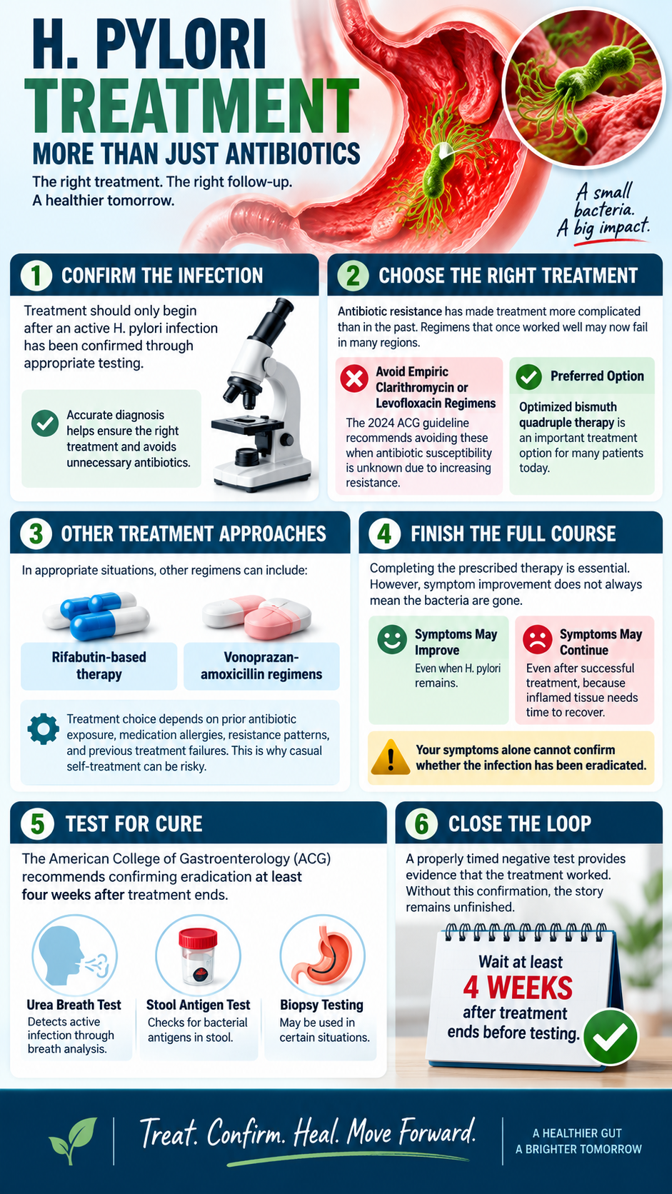
Other approaches can include rifabutin-based therapy or vonoprazan-amoxicillin regimens in appropriate situations. Treatment choice depends on prior antibiotic exposure, medication allergies, resistance patterns, and previous treatment failures. This is one reason casual self-treatment becomes risky when dealing with a persistent organism.
Completing therapy also does not automatically prove eradication. Symptoms can improve even when bacteria remain. Conversely, symptoms can continue after successful treatment because inflamed tissue needs time to recover. A person’s subjective experience therefore cannot substitute for a proper test of cure.
The ACG recommends confirming eradication at least four weeks after treatment ends. Urea breath testing, stool antigen testing, or appropriate biopsy testing can provide that confirmation.
That follow-up step closes the loop. Treatment represents an attempt to remove the organism, while a properly timed negative test provides evidence that the attempt succeeded. Without that confirmation, the story remains unfinished.
Where Functional Support Can Add Value
Eradicating H. pylori solves one important problem, but it does not automatically reverse years of gastric dysfunction. Chronic inflammation may leave the stomach lining irritated, nutrient status altered, and digestive function compromised. Antimicrobial therapy can also affect microbial communities throughout the gastrointestinal tract.
That is where thoughtful functional support can become useful. The goal should involve strengthening recovery, improving treatment tolerance, correcting deficiencies, and supporting normal digestion. It should not involve replacing proven eradication therapy with a large collection of supplements and hoping the organism disappears.
Probiotics provide one useful example. A large systematic review and network meta-analysis examined 91 randomized controlled trials involving 13,680 participants. Adding probiotics to standard treatment improved eradication outcomes and reduced several treatment-related side effects. Diarrhea, nausea, bloating, and abdominal discomfort occurred less often in many probiotic groups.
Those findings do not mean probiotics reliably eliminate H. pylori by themselves. Their value appears strongest as supportive tools used alongside appropriate treatment. That distinction allows conventional eradication and functional recovery to work together rather than competing as opposing philosophies.
The body responds to physiology, not ideology. Removing the pathogen matters, and rebuilding the environment it damaged matters too. A complete strategy recognizes both goals instead of pretending only one deserves attention.
Rebuilding the Gastric Terrain After Eradication
After eradication, the next phase involves restoring the stomach’s ability to function normally. Tissue healing requires adequate protein, micronutrients, blood flow, sleep, and time. A damaged gastric lining cannot rebuild itself from highly processed food and nutrient-poor calories alone.
Protein provides amino acids used in tissue repair and enzyme production. Zinc supports immune function and epithelial healing, while vitamins A and C contribute to tissue integrity and antioxidant defense. Vitamin B12, iron, folate, and other nutrients may require direct evaluation when chronic gastritis has been present for years.
Whole-food nutrition becomes especially valuable during recovery. Highly processed foods often deliver excessive refined carbohydrates, damaged oils, additives, and very little micronutrient density. A nutrient-rich diet provides better material for rebuilding while reducing some of the dietary exposures associated with poor gastric health.
Individual tolerance should guide the transition. Some people tolerate raw vegetables and acidic foods easily, while others find them irritating during active gastritis. Large meals may worsen pressure and discomfort in a sensitive stomach, while smaller meals may feel more manageable. Healing works best when the diet responds to physiology rather than rigid ideology.
Smoking deserves particular attention because it compounds gastric risk. The National Cancer Institute reports higher stomach cancer risk among smokers infected with H. pylori. High salt intake and processed meats also associate with greater gastric cancer risk. Reducing those exposures therefore supports both short-term recovery and long-term gastric health.
What You Can Begin Doing at Home
Home care should begin with observation rather than a shopping cart full of supplements. Pay attention to the timing and pattern of symptoms. Notice whether discomfort appears before eating, immediately after meals, or several hours later. Track persistent nausea, belching, early fullness, abdominal burning, appetite changes, fatigue, and unusual stool changes.
Those observations do not diagnose H. pylori, but they create useful clinical information. Patterns can help determine whether testing deserves attention and whether other gastrointestinal problems may be present. A detailed symptom history often reveals clues that isolated complaints never show.
Medication history deserves the same scrutiny. Frequent NSAID use can aggravate stomach injury, while long-term acid suppression can influence both gastric physiology and H. pylori testing. Necessary medications should never be stopped casually, but every long-term prescription deserves a clear reason for continued use.
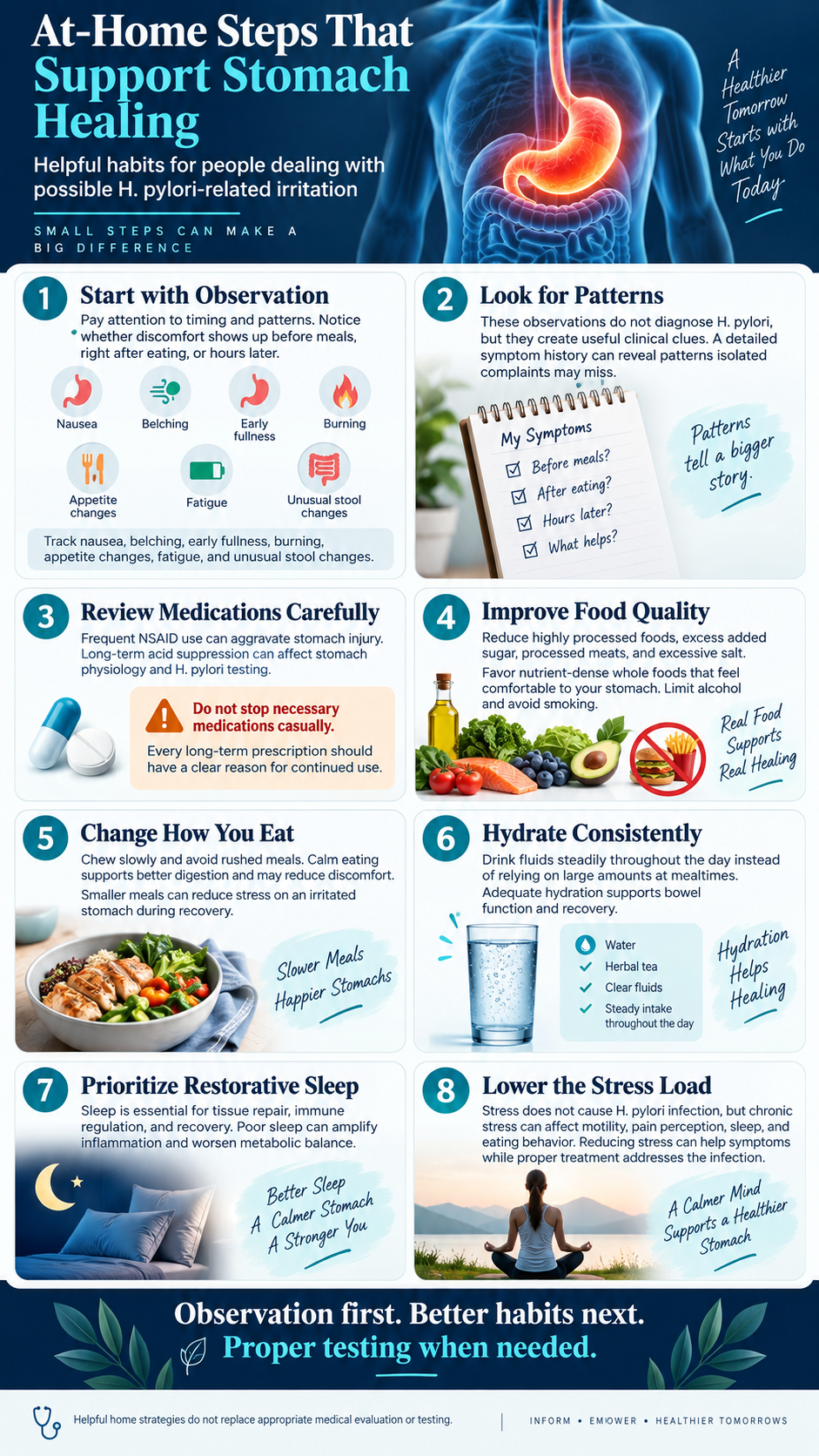
Food quality can also change the inflammatory burden. Reducing highly processed foods, excessive added sugar, processed meats, and excessive salt creates a better nutritional environment. Smoking should stop, and alcohol should remain limited when the stomach remains irritated. Meals should emphasize nutrient-dense whole foods that the person tolerates comfortably.
Eating behavior matters more than most people realize. Chewing slowly allows digestion to begin properly before food reaches the stomach. Rushed meals often encourage overeating, poor chewing, and greater digestive discomfort. A calmer meal environment can also support better parasympathetic activity and more coordinated digestion.
Meal size can become useful during active gastritis because enormous meals stretch an irritated stomach. Smaller portions may temporarily reduce discomfort until inflammation settles. The goal is not permanent food restriction, but rather reducing unnecessary mechanical stress during recovery.
Hydration supports normal physiology, although excessive fluid intake during meals is unnecessary. Most people benefit from drinking consistently throughout the day instead of relying on large volumes at mealtimes. Adequate hydration also supports bowel function and overall metabolic recovery.
Sleep deserves equal attention because tissue repair and immune regulation depend heavily on restorative sleep. Chronic sleep deprivation amplifies inflammatory signaling and worsens metabolic regulation. A healing digestive system works better inside a body receiving enough recovery time.
Stress requires a careful distinction. Psychological stress does not create H. pylori infection, but chronic stress can affect motility, pain perception, sleep, eating behavior, and autonomic balance. Reducing that physiological load can improve symptoms while the true infection receives appropriate treatment.
Supporting the Gut During Treatment
Eradication therapy can challenge the gastrointestinal system. Some patients develop nausea, diarrhea, metallic taste, abdominal discomfort, or bloating. These side effects can reduce treatment adherence and increase the chance of missed doses.
Selected probiotics may help reduce these problems. Current evidence suggests certain probiotic strategies can improve treatment tolerance and modestly increase eradication success. However, strains and formulations differ widely between studies, so support should remain individualized rather than generic.
Fermented foods can also support microbial diversity when tolerated. Unsweetened fermented vegetables and other traditional foods may provide beneficial organisms and metabolites. People with histamine intolerance or active gastrointestinal sensitivity may need a slower approach.
Prebiotic fibers can help beneficial intestinal bacteria recover after antimicrobial therapy. However, suddenly adding large amounts of fiber can worsen bloating in a sensitive digestive system. Gradual progression usually makes more physiological sense than forcing aggressive supplementation.
Investigate Nutrient Deficiencies Instead of Guessing
Longstanding gastric inflammation creates another important opportunity for investigation. Iron deficiency deserves particular attention because H. pylori can contribute through several mechanisms. Someone may repeatedly receive iron supplements without anyone asking why iron stores remain low.
Vitamin B12 can become equally important when extensive gastritis affects normal gastric physiology. Folate, magnesium, and other nutrients may also warrant evaluation depending on symptoms, diet, and laboratory findings. The principle remains simple: test what matters instead of guessing blindly.
Correcting a documented deficiency can improve energy, cognition, blood production, and overall recovery. However, supplementation works best when the underlying cause has also been addressed. Replacing iron while ignoring chronic gastric inflammation can become an endless cycle of temporary improvement.
A more complete strategy asks two questions at once. What nutrient is low, and why did it become low? That second question often reveals the deeper mechanism.
Know When Home Care Is Not Enough
At-home strategies should support medical evaluation rather than delay it. Black stools may indicate gastrointestinal bleeding, while vomiting blood requires immediate attention. Persistent vomiting, unexplained weight loss, progressive swallowing difficulty, or severe abdominal pain also deserve prompt evaluation.
Worsening anemia should never become a supplement experiment. These symptoms can signal ulcers, bleeding, obstruction, malignancy, or another significant gastrointestinal disorder. Proper investigation becomes more important as warning signs increase.
Functional thinking does not mean rejecting conventional diagnostics. It means using diagnostics to uncover mechanisms instead of merely labeling symptoms. Endoscopy, imaging, pathology, and laboratory testing can become powerful tools when used for the right clinical question.
The Test That Closes the Case
One final step separates complete H. pylori care from unfinished care: retesting after treatment. Symptoms cannot confirm eradication because they may improve before the organism disappears. They can also remain after successful eradication because the tissue still needs time to heal.
Current guidelines recommend confirming eradication after therapy. Testing should occur at least four weeks after treatment ends. PPIs generally require a two-week hold beforehand, while antibiotics and bismuth require four weeks.
A properly timed negative test changes the entire clinical conversation. At that point, persistent symptoms deserve a new investigation rather than another blind round of antimicrobial therapy. The original infection may be gone while inflammation, motility problems, dysbiosis, or another condition remains.
Closure requires evidence. Feeling better is encouraging, but confirmation provides certainty. That principle becomes especially important with an organism known for surviving silently.
The Deeper Lesson of H. pylori
The story of H. pylori reveals something larger than a single bacterial infection. It shows how easily symptoms can distract attention from mechanisms. Burning gets labeled as an acid problem, fatigue becomes an iron problem, bloating becomes food intolerance, and nausea becomes a medication side effect.
Each symptom may receive its own treatment while the connecting cause stays buried underneath. Sometimes that cause has been living inside the stomach since childhood. Years of chronic inflammation can then reshape digestion long before anyone recognizes the pattern.
H. pylori survives because it adapts brilliantly to human physiology. It creates a protective chemical environment, travels through gastric mucus, attaches to the stomach lining, and avoids complete immune destruction. The price of that survival is chronic inflammation within a tissue designed to perform delicate digestive functions.
Over time, the consequences can extend far beyond occasional indigestion. Ulcers may develop, iron stores may fall, acid production may change, and gastric tissue may become atrophic. In susceptible people, intestinal metaplasia and malignancy can eventually enter the picture.
The progression is neither inevitable nor harmless. Early recognition creates an opportunity to interrupt that process before deeper damage develops. That possibility makes intelligent testing and complete eradication especially important.
The most powerful response begins with curiosity rather than assumption. Ask what created the symptom, choose the right test, remove confirmed infection, verify eradication, and rebuild the environment that chronic inflammation disrupted. That sequence transforms care from symptom management into genuine investigation.
The human body continually attempts to maintain balance and repair damaged tissue. Healing becomes more difficult when an ongoing infection keeps provoking injury. Removing that obstacle can create the biological conditions needed for repair to proceed more effectively.
For some people, H. pylori entered the stomach before they were old enough to remember becoming sick. Decades later, finally identifying it may explain a story their symptoms have been trying to tell for years.