
The Inflammation Hiding Beneath the Surface
Inflammation has a public-relations problem. Most people picture something obvious when they hear the word: a swollen knee, an infected cut, or a painful joint. Those are visible examples of inflammation doing exactly what biology designed it to do. Yet another form can develop quietly, without redness, swelling, fever, or obvious pain. This persistent inflammatory activity may accompany metabolic dysfunction, cardiovascular disease, autoimmune conditions, chronic infections, obesity, and other disorders. Someone can feel relatively normal while inflammatory biology continues beneath the surface. That possibility raises an important question: how do we detect inflammation that does not announce itself?
For decades, clinicians have relied on markers such as C-reactive protein, erythrocyte sedimentation rate, and white blood cell counts. These tests remain useful, but they examine inflammation through particular biological windows. None captures every component of the inflammatory response. More advanced laboratory technology has introduced another marker that approaches the problem differently. It is called GlycA, and despite its growing research history, most patients have never heard its name.
GlycA deserves attention because it does not simply measure another isolated inflammatory protein. Instead, it reflects a composite signal arising from several circulating glycoproteins involved in the acute-phase response. That distinction offers a broader view of inflammatory activity. More importantly, an abnormal GlycA result should provoke a deeper question. The question is not simply, “How do we lower this number?” It is, “Why is the body producing this inflammatory signal?”
What Is GlycA?
GlycA stands for glycoprotein acetylation. Laboratories measure it using nuclear magnetic resonance spectroscopy, commonly called NMR. The same technology can characterize lipoprotein particles and other circulating molecules. With GlycA, NMR detects signals from N-acetyl methyl groups attached to certain glycoproteins circulating within the blood.
Several acute-phase proteins contribute substantially to the GlycA signal. These include alpha-1-acid glycoprotein, haptoglobin, alpha-1-antitrypsin, and other glycosylated proteins. The liver produces many of these proteins as part of the body’s inflammatory response. Their concentrations and glycosylation patterns can change when immune signaling increases.

This makes GlycA fundamentally different from a test measuring one molecule. It represents the combined behavior of several proteins associated with inflammatory physiology. Think of the difference between listening to one instrument and hearing an entire section of an orchestra. One instrument provides useful information, but several instruments reveal something about the larger performance.
That does not make GlycA a diagnostic test for one particular disease. An elevated result cannot tell someone that they have diabetes, cardiovascular disease, an autoimmune disorder, or an infection. Instead, it signals that inflammatory biology may be more active than expected. The next step is determining why.
GlycA and CRP Tell Different Parts of the Story
C-reactive protein, particularly high-sensitivity CRP, has become one of medicine’s most recognized inflammatory biomarkers. The liver produces CRP in response to inflammatory cytokines, especially interleukin-6. Levels can rise dramatically during infection, trauma, or other acute inflammatory events. Smaller elevations can also appear alongside chronic cardiometabolic disease.
GlycA approaches inflammation from another direction. Rather than quantifying one protein, it integrates signals from several acute-phase glycoproteins. Those proteins possess different circulating half-lives and respond differently to biological stress. Consequently, GlycA may provide information that overlaps with CRP without merely duplicating it.
The important point is not that GlycA should replace CRP. Medicine rarely becomes stronger by exchanging one isolated number for another. The greater value comes from patterns. GlycA, hs-CRP, fasting insulin, glucose, hemoglobin A1c, triglycerides, blood counts, liver markers, and other appropriate tests can reveal different pieces of physiology.
When several markers move in the same unfavorable direction, the biological picture becomes harder to dismiss. Conversely, conflicting results may encourage a more careful investigation. Laboratory medicine works best when numbers create questions rather than prematurely ending them.
Inflammation Is Not the Enemy
Before discussing chronic inflammation, we need to correct a common misunderstanding. Inflammation is not inherently harmful. Without an inflammatory response, humans could not survive ordinary infections or repair damaged tissue.
Imagine cutting your finger. Blood vessels change, platelets respond, immune cells arrive, and chemical messengers coordinate repair. The affected area becomes warm, swollen, and tender because the body has launched an organized defense. Once the threat passes, specialized processes help resolve the inflammatory response and restore tissue stability.
The problem begins when inflammatory signaling fails to resolve appropriately or receives repeated stimulation. Instead of a short emergency response, the body enters a prolonged state of biological defense. The signal may remain relatively quiet, yet its cumulative effects can become significant.
Chronic inflammation is therefore better understood as an environment than as a single event. Immune cells, liver proteins, adipose tissue, hormones, blood vessels, mitochondria, and metabolic pathways can all participate. GlycA offers one window into this larger biological conversation.
Where Does Chronic Inflammation Come From?
There is rarely one universal cause. Chronic inflammatory activity can emerge from several overlapping sources, and identifying those sources matters more than simply naming inflammation.
Metabolic dysfunction deserves particular attention. Insulin resistance can develop long before fasting glucose reaches the diabetic range. The pancreas initially compensates by releasing additional insulin, helping maintain apparently acceptable glucose levels. Meanwhile, metabolic stress can continue building beneath the surface.
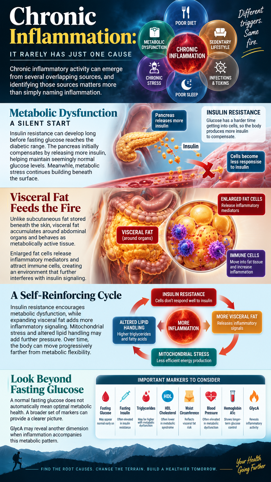
Visceral fat can intensify this process. Unlike subcutaneous fat stored beneath the skin, visceral adipose tissue accumulates around abdominal organs and behaves as metabolically active tissue. Enlarged fat cells can release inflammatory mediators while attracting immune cells into adipose tissue. This inflammatory environment can further interfere with insulin signaling.
A self-reinforcing cycle may then emerge. Insulin resistance encourages metabolic dysfunction, while expanding visceral fat contributes additional inflammatory signaling. Mitochondrial stress and altered lipid handling may add further pressure. Over time, the body can move progressively farther from metabolic flexibility.
That is one reason a normal fasting glucose does not automatically prove optimal metabolic health. Fasting insulin, triglycerides, HDL cholesterol, waist circumference, blood pressure, and hemoglobin A1c can add valuable context. GlycA may reveal another dimension when inflammation accompanies that metabolic pattern.
The Gut Can Become Part of the Inflammatory Conversation
Another potential source sits inside the gastrointestinal tract. The intestinal lining performs an extraordinary balancing act. It must absorb nutrients while maintaining a barrier between the bloodstream and enormous numbers of microorganisms, food compounds, and microbial products.
A healthy intestinal barrier depends upon epithelial cells, mucus, immune defenses, tight-junction proteins, and a diverse microbial ecosystem. Disturbances in this system can alter local and systemic immune signaling. Gastrointestinal infections, inflammatory bowel disease, alcohol misuse, medications, dietary factors, and metabolic disease can influence this environment.
Researchers continue investigating intestinal permeability and microbiome changes in chronic inflammatory disorders. The science is promising but complicated. Not every symptom represents “leaky gut,” and an elevated GlycA result cannot prove that intestinal permeability caused systemic inflammation.
However, persistent digestive symptoms should not be ignored when inflammatory markers remain elevated. Chronic diarrhea, constipation, bloating, abdominal pain, unexplained nutrient deficiencies, or previous gastrointestinal disease may justify deeper investigation. The goal is not blaming every illness on the gut. The goal is recognizing that intestinal and immune physiology are intimately connected.
The Liver Sits at the Center of the GlycA Story
The liver deserves special attention because it produces many proteins contributing to the GlycA signal. During inflammatory stress, cytokines communicate with liver cells and alter protein production. This acute-phase response changes concentrations of several circulating proteins involved in immunity, transport, coagulation, and tissue protection.
Metabolic dysfunction can simultaneously place tremendous pressure on the liver. Insulin resistance and excess energy availability can promote hepatic fat accumulation. Metabolic dysfunction-associated steatotic liver disease can develop silently, sometimes before obvious symptoms appear.
This creates an important intersection between metabolism and inflammation. The liver does not merely process nutrients. It helps coordinate glucose regulation, lipid metabolism, detoxification pathways, protein production, and immune signaling. When metabolic health deteriorates, hepatic physiology can become part of a much larger systemic problem.
Standard liver enzymes can provide useful information, but they do not describe every aspect of liver health. Clinical history, metabolic markers, alcohol exposure, medications, body composition, and imaging may provide additional context when indicated. GlycA cannot diagnose liver disease, yet its biology reminds us how closely inflammation and hepatic function interact.
What Persistent Inflammation Can Do to the Body
Chronic inflammatory activity matters because the effects can extend far beyond one organ. Blood vessels contain an active endothelial lining that regulates vascular tone, clotting, immune interactions, and nutrient exchange. Metabolic and inflammatory stress can impair endothelial function and contribute to conditions that support atherosclerosis.
The brain also participates in inflammatory signaling. Immune mediators communicate with neural pathways and can influence energy, appetite, sleep, and cognition. This interaction helps explain why acute illness often produces fatigue and mental sluggishness. Persistent inflammation may contribute to similar complaints in some chronic conditions, although fatigue and brain fog always require a broader differential evaluation.

Skeletal muscle can also become metabolically resistant to insulin, reducing efficient glucose disposal. Adipose tissue may become increasingly inflammatory. The liver can accumulate fat. Blood vessels can experience oxidative and inflammatory stress. None of these processes occurs completely independently.
This interconnected biology explains why GlycA becomes so interesting. The test is not important because one laboratory number holds the secret to disease. It matters because it can expose a biological pattern that deserves investigation.
The real story of GlycA is therefore not the marker itself. The story is what may be generating the signal. When chronic inflammation appears, the most valuable question is not how to silence the laboratory alarm. The better question asks where the biological fire began, what continues feeding it, and what must change so the body can finally move from defense toward repair.
WHEN CHRONIC INFLAMMATION BECOMES A WHOLE-BODY PROBLEM
The most important lesson from GlycA begins after an elevated result appears. Finding inflammation is only the first step. The real work involves understanding what sustained inflammatory signaling may be doing throughout the body. Chronic inflammation rarely respects anatomical boundaries. Metabolism influences the liver, the liver influences circulation, and blood vessels communicate with immune and endocrine systems. Meanwhile, skeletal muscle, adipose tissue, the gastrointestinal tract, and the brain participate in the same biological conversation. A problem that begins quietly can eventually become a network problem.
This distinction changes how we should think about inflammatory health. Lowering one laboratory marker cannot become the entire objective. The greater goal involves improving the physiological environment producing that marker. When metabolic stress decreases, sleep improves, muscles become more active, and excessive visceral fat declines, inflammatory signaling may also improve. That approach moves beyond chasing numbers and focuses attention on the conditions surrounding them.
The Cardiovascular Connection Goes Beyond Cholesterol
For decades, cardiovascular conversations have centered heavily on cholesterol. Lipoproteins certainly matter, but atherosclerosis involves considerably more biology than a cholesterol measurement alone can describe. The arterial wall is living tissue filled with endothelial cells, smooth muscle cells, immune cells, signaling molecules, and structural proteins. Inflammation participates throughout the development and progression of atherosclerotic disease.
Damage or dysfunction within the vascular environment can encourage immune activity. Certain lipoproteins can enter and become retained within the arterial wall. Immune cells respond to that environment, creating inflammatory activity inside developing lesions. Smoking, hypertension, diabetes, insulin resistance, and other factors can intensify the process. Cardiovascular disease therefore develops through interacting lipid, metabolic, vascular, and inflammatory pathways.
Research has associated higher GlycA concentrations with cardiovascular events in several populations. That relationship does not establish GlycA as the cause of heart disease. Instead, GlycA may capture inflammatory activity accompanying underlying cardiovascular risk. This distinction matters because biomarkers should guide investigation rather than become villains themselves.
A comprehensive cardiovascular assessment may therefore examine blood pressure, glucose regulation, smoking exposure, family history, lipoproteins, body composition, activity levels, and inflammatory markers. Some individuals may require additional testing based on their risk profile. Looking beyond a standard cholesterol panel can reveal biological problems that otherwise remain hidden.
Inflammation Can Disrupt Metabolic Communication
Every meal requires extraordinary coordination. The digestive tract absorbs nutrients, the pancreas adjusts hormone production, the liver manages incoming fuel, and skeletal muscle responds to insulin. Adipose tissue stores or releases energy depending upon hormonal signals. When these systems communicate efficiently, glucose and lipids move through predictable metabolic pathways.
Insulin resistance disrupts that coordination. Muscle cells may become less responsive to insulin, forcing the pancreas to compensate with greater insulin secretion. The liver may continue producing glucose when it should reduce output. Adipose tissue can release more fatty acids into circulation. These changes place increasing pressure on metabolic control.
Inflammatory signaling can become intertwined with this process. Certain inflammatory pathways can interfere with normal insulin signaling inside cells. Meanwhile, metabolic dysfunction can create additional oxidative and inflammatory stress. The relationship can therefore move in both directions rather than following one simple cause-and-effect pathway.
Eventually, compensation may become inadequate. Blood glucose rises more consistently, hemoglobin A1c increases, and type 2 diabetes can emerge. Long before that stage, however, someone may already have hyperinsulinemia, expanding visceral fat, abnormal triglycerides, or other warning signs. GlycA can add another layer when systemic inflammation accompanies those metabolic changes.
Chronic Inflammation Can Drain Energy
Fatigue often gets dismissed as a consequence of aging, stress, or a demanding schedule. Sometimes those explanations are correct. However, persistent exhaustion can also accompany metabolic, endocrine, sleep, infectious, hematologic, or inflammatory problems.
The immune system requires energy. During an infection, the body deliberately changes behavior and metabolism to prioritize defense. Appetite can decline, activity decreases, sleepiness increases, and concentration becomes more difficult. Scientists sometimes describe these coordinated changes as sickness behavior.
Persistent inflammatory signaling may produce subtler versions of those effects in certain conditions. At the same time, insulin resistance can make energy regulation less efficient. Poor sleep may worsen glucose control, while inadequate activity reduces mitochondrial demand. Multiple small disruptions can eventually produce a person who feels depleted despite consuming plenty of calories.
Fatigue should never automatically be blamed on inflammation. Anemia, thyroid disorders, sleep apnea, medication effects, infections, nutrient deficiencies, depression, and numerous diseases can produce similar symptoms. The important lesson involves refusing to normalize persistent exhaustion without investigating plausible causes.
The Brain Is Listening to the Immune System
The brain and immune system communicate constantly. Cytokines can influence neural activity, while the autonomic nervous system helps regulate immune responses. Hormones add another layer to this conversation. The result is a sophisticated network connecting stress, immunity, metabolism, behavior, and neurological function.
Inflammatory signaling can influence concentration, motivation, sleep, and mood during illness. Anyone who has experienced influenza understands the phenomenon. The brain feels slower because the body has shifted resources toward recovery.
Chronic conditions can produce more complicated versions of this interaction. Some patients describe mental fog, poor concentration, reduced stamina, or disrupted sleep alongside inflammatory disease. These symptoms remain nonspecific and should never serve as proof of elevated GlycA.
Nevertheless, they reinforce an important concept. Systemic inflammation is not confined to the location where it originated. Chemical messengers travel. Hormones respond. Neural pathways react. The body behaves as an integrated system.
Why Suppressing the Signal Is Not Enough
Once someone discovers elevated inflammation, the temptation is to search for the fastest anti-inflammatory supplement. Curcumin, omega-3 fatty acids, antioxidants, herbs, and countless formulations dominate the wellness marketplace. Some compounds have legitimate research behind them, but supplementation cannot replace identifying major inflammatory drivers.
Imagine smoke filling a kitchen because food is burning on the stove. Opening a window might reduce the smoke. The intervention helps, but the pan remains on the burner. Effective problem-solving requires removing the source.
The same principle applies to inflammatory physiology. Someone cannot out-supplement persistent smoking, severe sleep deprivation, uncontrolled diabetes, excessive alcohol intake, significant visceral obesity, or an untreated inflammatory disease. Supplements may complement a broader strategy when appropriate. They should not distract from foundational problems.
The first objective should therefore involve identifying the largest modifiable contributors. Once those factors become visible, daily behavior becomes one of the most powerful places to begin.
The At-Home GlycA Strategy: Start With Food Quality
Nutrition offers an immediate opportunity to change metabolic inputs. The objective is not starvation or an elaborate detoxification program. The goal involves reducing foods that encourage overconsumption while emphasizing nutrient-dense meals that support satiety and metabolic control.
Begin by dramatically reducing sugar-sweetened beverages, refined snacks, desserts, and heavily ultra-processed foods. These products can make excessive energy consumption remarkably easy. Replace them with meals centered around adequate protein and minimally processed ingredients. Eggs, fish, poultry, meat, vegetables, avocados, olives, and other tolerated whole foods can provide useful building blocks.

Carbohydrate needs differ considerably among individuals. Someone with substantial insulin resistance may respond differently than an endurance athlete. Instead of treating every carbohydrate as identical, consider quantity, processing, glycemic response, activity level, and metabolic status. Food should serve physiology rather than ideology.
Meal frequency also deserves attention. Constant grazing creates repeated opportunities for excessive intake. Many adults benefit from defined meals and fewer unnecessary snacks. Appropriate overnight fasting can naturally create a period without incoming calories. Anyone using glucose-lowering medication should discuss significant dietary or fasting changes with the prescribing clinician.
Make Skeletal Muscle Part of the Treatment Strategy
Muscle is one of the body’s largest destinations for glucose. When muscle contracts, glucose uptake can increase through mechanisms that complement insulin signaling. Building and regularly using muscle therefore provides a powerful tool for metabolic health.
Resistance exercise two or more times weekly can improve strength, preserve lean mass, and support insulin sensitivity. Training does not require bodybuilding equipment. Squats, lunges, push-ups, resistance bands, dumbbells, and appropriately challenging bodyweight movements can create meaningful muscular demand.
Walking adds another remarkably accessible intervention. Ten to fifteen minutes of movement following meals can help reduce post-meal glucose excursions. A longer daily walk can increase energy expenditure while improving cardiovascular fitness and stress regulation.
The key is frequency. One punishing workout cannot compensate for a week spent almost entirely seated. Build movement into everyday life until muscular activity becomes normal rather than exceptional.
Treat Sleep Like a Metabolic Requirement
Poor sleep can undermine nearly every other health strategy. Sleep restriction influences insulin sensitivity, appetite regulation, sympathetic nervous system activity, and inflammatory signaling. Someone can eat carefully and exercise consistently while chronic sleep deprivation continues working against those efforts.
Create a predictable sleep schedule whenever possible. Keep the bedroom dark, quiet, and comfortably cool. Reduce stimulating screen exposure near bedtime and avoid using alcohol as a sleep aid. Morning outdoor light can help reinforce circadian timing and support a more predictable sleep-wake rhythm.
Persistent snoring, witnessed breathing pauses, morning headaches, or severe daytime sleepiness require more than sleep hygiene. Those symptoms can accompany obstructive sleep apnea. Addressing an underlying sleep disorder may produce substantially greater benefits than adding another supplement.
Reduce the Inflammatory Load You Can Control
Certain exposures deserve direct attention because no nutritional strategy makes them harmless. Cigarette smoking promotes oxidative stress, vascular injury, and inflammatory activity. Excessive alcohol can impair sleep, damage the liver, alter gastrointestinal function, and complicate metabolic health.
Stress also matters, although telling someone to “reduce stress” accomplishes little. Create daily periods that deliberately shift the nervous system away from constant stimulation. Walking outdoors, prayer, meditation, slow breathing, social connection, and quiet time can provide practical opportunities for recovery.
Environmental perfection is impossible. Focus first on high-impact exposures rather than becoming frightened of every chemical encountered during normal life. Improving ventilation, avoiding tobacco smoke, using appropriate workplace protection, and maintaining safe drinking water provide sensible foundations.
Recheck the Biology Instead of Guessing
After several months of consistent changes, reassessment can reveal whether the internal environment is actually improving. Depending upon individual circumstances, this may include GlycA alongside hs-CRP, fasting glucose, fasting insulin, hemoglobin A1c, triglycerides, HDL cholesterol, blood pressure, waist circumference, and relevant liver markers.
The objective is not achieving a beautiful laboratory report while ignoring how the person functions. Better metabolic health should eventually appear across several domains. Waist circumference may decrease, blood pressure can improve, energy may become steadier, glucose regulation can strengthen, and inflammatory markers may move favorably.
If GlycA remains substantially elevated despite meaningful lifestyle improvements, that information matters. Persistent inflammation can justify investigating other sources, including infection, autoimmune disease, inflammatory disorders, liver disease, or other medical conditions. Root-cause thinking requires enough humility to keep investigating when the first explanation fails.
GlycA ultimately offers something more valuable than another number on a laboratory report. It offers a reason to look beneath the surface. Chronic inflammation often develops through years of accumulated metabolic, environmental, immune, and lifestyle pressures. Those forces can affect arteries, metabolism, energy production, liver function, and neurological signaling long before one dramatic symptom provides a warning.
The purpose of measuring inflammation should never be fear. It should be information followed by intelligent action. Find the likely sources. Change the factors that can be changed. Investigate the ones that cannot be explained. Then measure again and allow the biology to reveal whether the strategy is working.
The body does not need another attempt to silence its warning lights. It needs us to understand why those warning lights came on in the first place.