
The Pain in Your Back May Begin Beneath Your Feet
The human body rarely develops chronic pain without a history. Sometimes that history begins with an injury, repetitive strain, or years of physical inactivity. Other times, the story starts somewhere few people think to investigate: the soles of their feet. A person may spend years treating lower back pain while overlooking mechanical problems that influence every step.
Imagine a man who wakes each morning with stiffness across his lower back. He stretches, changes mattresses, visits specialists, and occasionally finds relief through spinal treatment. Yet the discomfort returns whenever he spends several hours walking or standing. Nobody has examined his ankle mobility, foot stability, or the way his body distributes weight.
His story illustrates a possibility that deserves more attention in conversations about chronic lower back pain. The painful region does not always reveal every contributing factor. Muscles, joints, connective tissues, and the nervous system work together to maintain movement. When one region struggles, neighboring structures may compensate, sometimes creating additional mechanical demands.
Your feet represent the body’s primary connection to the ground during standing and walking. Every step requires them to absorb loading, maintain stability, and help propel your body forward. When foot mechanics change, the effects can extend through the ankles, knees, hips, and pelvis. Under certain circumstances, these changes may influence the forces acting on the lumbar spine.
This relationship offers an important lesson about musculoskeletal health. Treating the location of pain matters, but understanding the movement system surrounding it matters too. Chronic back pain often involves several interacting factors rather than one defective joint. Examining the feet may reveal an overlooked contributor to a much larger problem.
The Human Foot: An Extraordinary Foundation Under Constant Pressure
Leonardo da Vinci reportedly described the human foot as an engineering masterpiece, and its anatomy supports that admiration. Each foot contains 26 bones, 33 joints, and numerous muscles, tendons, ligaments, and sensory receptors. Together, these structures create a foundation capable of adapting to unpredictable surfaces while supporting the body’s weight.
Unlike a concrete foundation, however, the foot must continually change shape during movement. Its arches flatten slightly under loading, then recover as weight shifts toward the toes. Small muscles help control these movements, while tendons and ligaments store and release mechanical energy. This remarkable coordination allows humans to walk efficiently across many different surfaces.
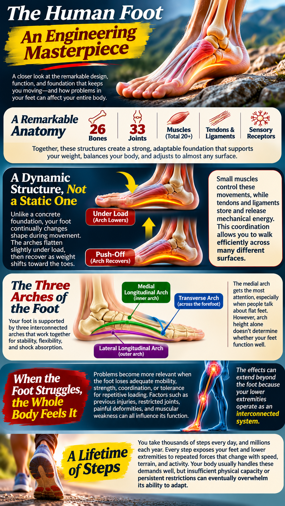
Three arches contribute to this process: the medial longitudinal, lateral longitudinal, and transverse arches. The medial arch attracts the most attention because people associate its appearance with flat feet. Yet arch height alone cannot determine whether someone has healthy foot mechanics. A flexible, relatively flat foot may function exceptionally well throughout an entire lifetime.
Problems become more relevant when the foot loses adequate mobility, strength, coordination, or tolerance for repetitive loading. Previous injuries, restricted joints, painful deformities, and muscular weakness can all influence its function. The consequences may extend beyond localized discomfort because the lower extremities operate as interconnected mechanical systems.
Consider the enormous number of steps accumulated throughout an ordinary week. Each movement exposes the feet and lower extremities to repeated forces that change with speed, terrain, and activity. The body usually handles these demands remarkably well, even when movement patterns differ between individuals. However, insufficient physical capacity or persistent painful restrictions may eventually overwhelm a person’s ability to adapt.
The Kinetic Chain: How Foot Problems Can Influence the Spine
Biomechanics describes the connection between the feet and spine through the kinetic chain. This concept recognizes that movement at one joint can influence neighboring joints during weight-bearing activities. The foot interacts with the ground, while the ankle, knee, hip, and pelvis coordinate the body’s response. The lumbar spine participates in this process by helping stabilize and position the trunk.
During walking, the foot undergoes a natural movement called pronation. This coordinated action allows the foot to adapt to the ground and accept body weight. Pronation commonly accompanies internal rotation of the tibia during the early stance phase. The knee and hip then participate in a complex sequence of movements that supports forward progression.
When pronation becomes prolonged or poorly controlled, the timing of these movements may change. Some individuals demonstrate altered knee rotation, hip positioning, or pelvic motion during walking. Their spinal muscles may respond by changing how they stabilize the trunk. Repeated exposure to uncomfortable loading can contribute to symptoms in susceptible people.
Nevertheless, the relationship does not follow a rigid mechanical formula. A particular amount of foot pronation does not guarantee a corresponding amount of pelvic rotation. Individual anatomy, muscular coordination, walking speed, and joint mobility all influence the outcome. Consequently, foot mechanics deserve examination without automatically becoming the explanation for every case of back pain.
The real concern involves the body’s ability to manage forces efficiently and comfortably. When one region cannot provide sufficient movement or stability, other regions may assume additional demands. These compensations often work successfully, allowing people to remain active without discomfort. Problems arise when those demands exceed tissue capacity or contribute to persistent symptoms.
Overpronation: When the Foot’s Natural Movement Becomes Difficult to Control
Pronation itself is not a disease, deformity, or mechanical mistake. Healthy feet naturally pronate during walking because flexibility helps accommodate the ground. The concern develops when the movement’s timing, magnitude, or muscular control creates difficulties during specific activities. Excessive pronation may contribute to altered loading, although many people with pronated feet remain completely pain-free.
Imagine a foot that rolls inward substantially as body weight passes over it. The tibia may rotate inward alongside this movement, influencing the knee’s position during stance. Depending on hip strength and motor control, the femur and pelvis may also adjust. These changes can modify the mechanical demands placed upon the lower back.
Over time, some individuals develop muscular fatigue or discomfort during prolonged walking and standing. Others experience symptoms primarily around the ankles, knees, or hips rather than the spine. The location of pain depends on numerous factors, including previous injuries and tissue sensitivity. Therefore, identifying overpronation requires more than observing a low arch or worn shoe.
Scientific research provides an intriguing connection between dynamic foot function and lower back pain. The Framingham Foot Study found an association between pronated foot function and low back pain among women. However, the researchers did not establish that pronation directly caused spinal discomfort. Their findings also demonstrated the importance of examining movement rather than relying exclusively on static foot posture.
This distinction changes how foot-related back pain should be investigated. A foot may look relatively normal while standing but behave differently during walking. Conversely, someone with visibly flat feet may demonstrate excellent balance, strength, and movement control. The important question concerns function, not whether every arch matches an idealized anatomical picture.
High Arches and Supination: When the Foot Becomes Too Rigid
Not every mechanical problem involves a collapsing arch or excessive inward movement. Some people experience the opposite pattern, characterized by unusually high arches and reduced foot flexibility. These individuals may distribute greater pressure along the heel and outer forefoot. Their feet can struggle to accommodate uneven surfaces, particularly when structural rigidity limits normal movement.
Supination describes a coordinated movement that helps the foot become more stable during propulsion. Like pronation, it serves an essential purpose during normal walking. Difficulties may develop when excessive rigidity limits the foot’s ability to adapt under loading. The ankles and knees may then experience different forces as the body attempts to maintain balance.
Individuals with rigid, high-arched feet sometimes develop recurrent ankle sprains, localized pressure pain, or difficulty finding comfortable footwear. Some compensate by changing their stride or shifting weight toward other regions. Those adaptations may influence hip and trunk mechanics, especially during demanding physical activities. However, current research does not establish high arches as a consistent independent cause of chronic lower back pain.
The distinction between structural appearance and functional performance remains essential. A high arch does not automatically require correction, just as a low arch does not guarantee dysfunction. Clinical attention becomes more important when foot structure accompanies pain, instability, restricted mobility, or progressive deformity. Understanding these differences prevents unnecessary treatment while identifying individuals who genuinely need additional support.
Limited Ankle Mobility: The Hidden Restriction That Changes Everything Above It
Consider what happens when someone attempts a deep squat with stiff ankles. Their knees cannot advance comfortably over their feet, forcing the body to find another movement strategy. The heels may lift, the feet may turn outward, or the trunk may lean farther forward. Each adjustment changes how the hips and lower back participate in the movement.
Ankle dorsiflexion allows the shin to advance over the foot during weight-bearing activities. Walking, climbing stairs, descending hills, and squatting all require this important movement. Restrictions may originate from previous sprains, calf tightness, joint stiffness, or painful ankle conditions. Years of avoiding certain movements can also reduce functional mobility and confidence.
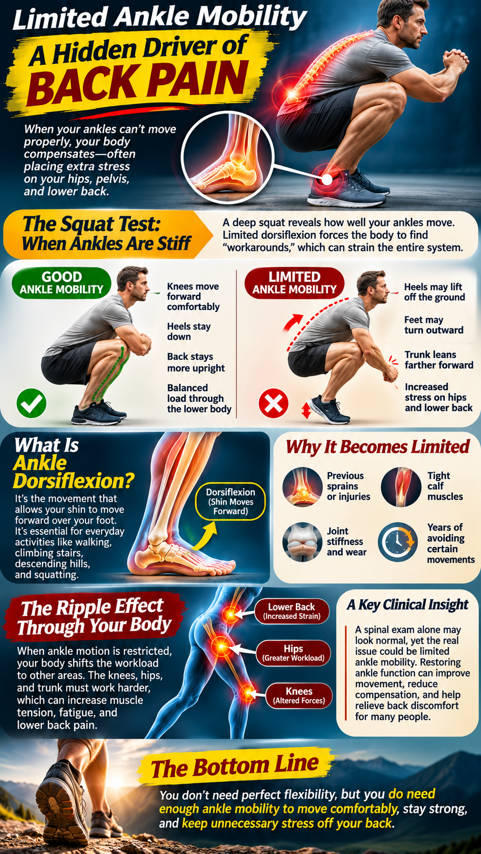
When dorsiflexion becomes limited, the body may redistribute movement demands toward the knees, hips, and trunk. These compensations can increase muscular effort during activities that require substantial ankle motion. For someone already experiencing lower back discomfort, the additional demands may aggravate symptoms. Nevertheless, the relationship depends on the activity and the individual’s movement strategy.
This explains why an examination limited to the lumbar spine can overlook useful information. A patient may demonstrate reasonable spinal mobility yet struggle with basic weight-bearing movements because of ankle restrictions. Improving ankle function may help restore more comfortable movement when the limitation contributes to symptoms. The objective is not perfect flexibility, but sufficient mobility for the person’s daily activities.
The Forgotten Big Toe and Its Influence on Walking Mechanics
Few people associate their big toe with lower back pain, yet this small joint influences an important walking phase. As the heel rises, the big toe normally extends while the body advances over the forefoot. This movement helps transform the foot into a stable lever for propulsion. Restricted motion can force a person to change how they complete each step.
The plantar fascia contributes through a mechanism known as the windlass effect. Big-toe extension increases tension along this connective tissue, supporting the foot’s longitudinal arch during push-off. When the joint becomes stiff or painful, normal propulsion may become less efficient. Arthritis, previous injuries, and structural deformities can all interfere with comfortable toe movement.
A person with restricted big-toe extension may shorten their stride or rotate the foot outward. Others shift pressure toward the smaller toes to avoid painful loading. These strategies can influence ankle and hip movement during walking. Over time, altered gait may increase discomfort elsewhere, although direct links to chronic back pain remain incompletely established.
This connection highlights the importance of examining the entire foot rather than focusing exclusively on arch height. The big toe, ankle, and plantar fascia contribute to coordinated movement through different mechanical functions. Limitations in any region may influence walking efficiency and physical comfort. Identifying those limitations creates opportunities for more individualized rehabilitation.
The Nervous System: How Your Feet Help Control Balance and Posture
The relationship between foot mechanics and back pain extends beyond bones, muscles, and joint angles. Thousands of sensory nerve endings help the feet detect pressure, movement, and contact with the ground. These signals travel through peripheral nerves toward the spinal cord and brain. The nervous system integrates this information to coordinate posture, balance, and movement.
Every step requires constant adjustments based on sensory feedback. Your brain combines information from the feet with signals from the eyes, muscles, joints, and vestibular system. Together, these inputs help determine where the body stands in space. Accurate sensory information allows the nervous system to respond efficiently to changing surfaces and movement demands.
When plantar sensation declines, balance and walking patterns may change significantly. Peripheral neuropathy illustrates this relationship because impaired sensation reduces awareness of ground contact. Some individuals respond by walking more cautiously or relying heavily on visual feedback. Others demonstrate increased instability, particularly when navigating darkness or uneven terrain.
Foot pain can also influence the nervous system’s movement strategies. Someone with a painful heel may unconsciously shorten their stance time on that side. A painful forefoot may encourage weight shifting or reduced push-off. These protective adaptations can modify movement throughout the lower extremities and trunk.
Importantly, the nervous system can also amplify pain without corresponding increases in tissue damage. Persistent pain may involve heightened sensitivity, altered movement confidence, and changes in muscular coordination. This helps explain why imaging findings and pain severity frequently disagree. Effective care must consider neurological processing alongside mechanical function rather than assuming every painful movement indicates structural injury.
The Pelvis and Lower Back: Where Mechanical Compensation Can Become Chronic Pain
The pelvis connects the lower extremities with the spine, making it central to walking and weight transfer. During each stride, it rotates and shifts while the trunk maintains balance. The hip muscles coordinate these movements, particularly during single-leg support. When the feet or ankles move differently, the pelvis may adjust its motion accordingly.
The gluteus medius helps control pelvic position when one foot supports the body’s weight. Meanwhile, the gluteus maximus contributes to hip extension and movement control during demanding activities. Weakness or insufficient endurance may alter how these muscles manage repetitive loading. The lumbar musculature may then assume additional demands during certain movements.
These adaptations do not necessarily indicate damage or spinal misalignment. Human movement naturally includes asymmetry, variation, and compensation. However, repeated activities can become uncomfortable when muscular capacity fails to match physical demands. Pain may gradually reduce activity, contributing to further weakness and declining movement tolerance.
A cycle can develop as discomfort encourages avoidance of walking, exercise, and prolonged standing. Reduced activity may weaken muscles that support efficient movement and decrease cardiovascular conditioning. As physical capacity declines, previously manageable activities become increasingly demanding. The resulting discomfort can reinforce inactivity and prolong the original problem.
This cycle helps explain why treating chronic back pain requires more than identifying one abnormal joint position. Foot function, hip strength, spinal mobility, conditioning, sleep, and pain sensitivity all deserve consideration. The most effective approach identifies which factors matter for the individual rather than assuming every patient shares the same cause.
The Missing Investigation in Chronic Back Pain
Modern imaging provides extraordinary insight into spinal anatomy, yet it cannot reveal every cause of musculoskeletal discomfort. An MRI may identify disc degeneration, joint changes, or narrowing around spinal nerves. However, many degenerative findings also appear in people who experience no pain. Imaging alone cannot explain how someone distributes weight or controls movement during daily activities.
A comprehensive examination should connect anatomical findings with symptoms and functional performance. Clinicians can evaluate walking patterns, ankle mobility, foot strength, balance, hip control, and spinal movement. These observations help identify potentially modifiable limitations that may contribute to discomfort. They also prevent unnecessary assumptions based solely on foot appearance or spinal imaging.
Emerging research supports investigating foot-related interventions in selected people with chronic lower back pain. A 2024 systematic review examined four randomized trials involving 268 participants with pronated feet and chronic back pain. The combined results suggested improvements in pain and disability following targeted physical interventions. However, the limited number of studies leaves uncertainty about which patients benefit most.
The larger message concerns clinical curiosity rather than promising a universal cure. Chronic back pain may involve spinal structures, muscular weakness, neurological sensitivity, or several interacting contributors. Foot mechanics can form part of that picture, especially when symptoms increase during weight-bearing activities. Ignoring the feet may leave a relevant source of mechanical stress unexplored.
Ultimately, understanding chronic lower back pain requires examining how the entire body functions during real movement. The feet create the initial contact with the ground, but the nervous system coordinates the response. Ankles, knees, hips, pelvis, and spine must work together to maintain comfortable movement. When that coordination becomes difficult, symptoms may appear far from the original limitation.
The path toward lasting improvement begins with identifying the factors that actually influence a person’s pain and physical function. For some, that investigation will reveal meaningful restrictions beneath the ankles. For others, the primary contributors will lie elsewhere in the movement system. Either way, examining the foundation offers a more complete understanding of the body above it.
Rebuilding the Foundation: A Functional Approach to Chronic Back Pain
A house with a shifting foundation rarely improves through repeated repairs to its walls. Contractors may patch cracks, replace damaged flooring, and repaint rooms until everything looks beautiful again. Yet the same problems return when the underlying instability remains unresolved. For some people with chronic lower back pain, their bodies follow a remarkably similar pattern.
Years of spinal discomfort often lead patients through an exhausting cycle of temporary relief and recurring symptoms. Massage may relax tight muscles, while chiropractic adjustments can improve comfort and mobility for certain individuals. Stretching sometimes reduces stiffness, and strengthening exercises may provide additional stability. However, meaningful improvement requires identifying the specific factors that continue placing excessive demands on the body.
Foot mechanics deserve attention because the feet influence movement throughout the lower extremities. Restricted ankles, painful toe joints, impaired balance, and inadequate muscular endurance can alter walking patterns. These changes may affect the knees, hips, pelvis, and lumbar spine during repetitive activities. Addressing relevant limitations can improve movement efficiency and potentially reduce discomfort in selected individuals.
A functional approach examines the body as an interconnected system rather than a collection of unrelated symptoms. It considers muscular strength, joint mobility, neurological coordination, and the demands of everyday life. The objective extends beyond temporarily reducing pain to restoring comfortable movement and physical resilience. Fortunately, many aspects of foot and lower-extremity function respond to consistent, appropriately designed home exercises.
The First Step Toward Healing: Understanding Your Walking Pattern
Most people rarely consider how they walk until pain begins interfering with ordinary activities. Walking appears automatic because the nervous system coordinates countless muscular contractions without conscious effort. Nevertheless, each stride requires precise timing between the feet, ankles, knees, hips, and trunk. Small movement differences may become clinically relevant when they coincide with persistent discomfort.
Begin by observing your walking pattern across a safe, level surface. Notice whether one foot turns outward, one heel rises earlier, or one stride feels shorter. Pay attention to discomfort that develops after several minutes rather than focusing exclusively on appearance. A short video from the side and behind may reveal patterns that are difficult to recognize independently.
Next, examine your standing posture without attempting to force your feet into an artificial position. Notice whether weight concentrates beneath the heels, forefeet, or outer edges. Observe differences between the right and left sides while keeping your knees relaxed. These observations provide useful information, although they cannot independently diagnose a biomechanical disorder.
Consider how symptoms respond to changes in activity, footwear, and walking duration. Pain that increases during prolonged standing may suggest different contributing factors than discomfort triggered by bending. Symptoms that consistently follow a particular movement deserve closer evaluation. Understanding these patterns helps guide rehabilitation toward meaningful functional improvements rather than unnecessary correction of harmless anatomical differences.
Strengthening the Foot’s Natural Support System
The foot contains small intrinsic muscles that help support its arches and control movement. These muscles work alongside larger muscles originating in the lower leg. Together, they contribute to stability, balance, and efficient weight transfer during walking. Like other skeletal muscles, they can improve their capacity through progressive exercise.
One useful exercise involves gently shortening the foot without curling the toes. While seated, place your foot flat against the floor and maintain relaxed toe contact. Draw the ball of the foot slightly toward the heel, creating a subtle arch lift. Hold the contraction for five seconds before relaxing and repeating approximately ten times.
This exercise, commonly called short-foot training, targets muscular control rather than forcing the arch into an exaggerated position. Beginners may find the movement surprisingly difficult because the required muscles often receive little conscious attention. Practice while seated before progressing to standing when control improves. Gradually introducing weight-bearing challenges helps the muscles adapt to more realistic demands.
Toe spreading offers another simple method for developing foot coordination. Place your feet flat on the floor and attempt to separate the toes without gripping the surface. Maintain the position briefly before relaxing and repeating several times. Although toe exercises cannot correct every structural deformity, they may improve muscular awareness and control.
Consistency matters more than intensity during the early stages of foot strengthening. A few minutes of focused practice most days can establish a foundation for progressive training. However, exercise should not produce escalating pain, numbness, or persistent swelling. Those symptoms warrant reassessment rather than forcing the foot through increasingly demanding movements.
Restoring Ankle Mobility to Reduce Unnecessary Compensation
Restricted ankle movement can influence how the body performs squats, climbs stairs, and walks across uneven terrain. When the ankle cannot move sufficiently, neighboring joints may compensate to complete the activity. Improving dorsiflexion may help restore comfortable movement when stiffness contributes to the problem. The key involves identifying restrictions and addressing them without aggravating existing injuries.
A practical home assessment uses the knee-to-wall movement. Stand facing a wall with one foot several inches away and the heel firmly grounded. Slowly bend the knee toward the wall while keeping it aligned with the toes. Move the foot closer or farther away to identify a comfortable distance that allows heel contact.
Differences between ankles may reveal limitations worth investigating, particularly when they accompany pain or functional difficulty. However, mild asymmetry does not automatically indicate dysfunction. Joint anatomy, previous injuries, and muscular flexibility all influence the available range. A clinician can help distinguish ordinary variation from restrictions that deserve treatment.
To improve calf flexibility, stand facing a wall and place one leg behind the other. Keep the rear heel grounded while gently leaning forward until the calf stretches comfortably. Hold the position for approximately thirty seconds before changing sides. Repeating the movement two or three times can provide a manageable starting routine.
A second variation targets the deeper soleus muscle by slightly bending the rear knee. This position changes the muscular emphasis while maintaining controlled ankle movement. Avoid bouncing or forcing the heel downward through pain. Gradual mobility training generally offers a more sustainable approach than aggressive stretching.
For individuals with previous ankle sprains, stiffness may involve joint restrictions rather than muscular tightness alone. Persistent swelling, instability, or painful limitations deserve professional evaluation. Proper rehabilitation may require strengthening, balance exercises, and progressive loading alongside mobility work. Restoring useful ankle function can improve movement throughout the lower extremities without promising automatic relief from spinal pain.
Reawakening the Big Toe and Improving Push-Off Mechanics
The big toe contributes significantly to the final phase of walking. As the heel rises, the toe extends and helps the foot develop a stable lever. Restricted motion may encourage shorter strides, altered pressure distribution, or compensatory foot rotation. Improving comfortable mobility can support more efficient walking when stiffness contributes to these changes.
Begin with gentle seated toe movements while keeping the foot relaxed. Carefully lift the big toe upward within a comfortable range before returning it to the floor. Repeat the movement slowly, avoiding pressure that causes sharp pain. Individuals with arthritis or significant joint deformities should avoid forcing motion beyond their comfortable limits.
Toe coordination exercises can also challenge the small muscles responsible for controlling forefoot movement. Try lifting the big toe while keeping the smaller toes against the ground. Then reverse the movement by lowering the big toe and gently raising the others. These exercises may initially feel awkward, but repetition often improves coordination.
Walking mechanics also depend on adequate calf strength and comfortable forefoot loading. Progressive heel raises can help develop the muscular capacity required for propulsion. Start with both feet grounded and use a countertop for balance. Slowly rise onto the balls of the feet before lowering the heels under control.
Begin with one or two sets of eight to twelve repetitions, depending on comfort and ability. Increase repetitions or resistance gradually as strength improves. People with painful toe arthritis, tendon injuries, or balance difficulties may require modified exercises. The objective is improved functional capacity rather than forcing every foot into an identical movement pattern.
Strengthening the Hips to Protect the Lower Back
Healthy foot mechanics cannot compensate for inadequate strength throughout the rest of the kinetic chain. The hips play a major role in controlling lower-extremity movement and supporting the pelvis. Their muscles help manage forces during walking, stair climbing, and single-leg activities. Weakness or insufficient endurance may increase demands on neighboring structures.
The gluteus medius contributes to pelvic control when one leg supports body weight. The gluteus maximus helps generate hip extension during standing, climbing, and lifting. Together, these muscles support efficient movement and reduce unnecessary reliance on other muscle groups. Improving their capacity may benefit people whose back pain accompanies reduced physical conditioning.
A simple starting exercise involves the glute bridge. Lie comfortably on your back with knees bent and feet resting on the floor. Gently tighten the abdominal muscles before lifting the hips without excessively arching the lower back. Hold briefly, then lower the pelvis with control.
Perform eight to twelve repetitions while maintaining comfortable breathing and movement. Progress gradually toward additional sets or longer holds as endurance improves. The exercise should create muscular effort without provoking significant spinal discomfort. Individuals with painful spinal conditions may need modifications based on their symptoms.
Side-lying hip abduction provides another option for strengthening muscles around the pelvis. Lie on one side with the lower knee bent for stability. Keep the upper leg relatively straight while lifting it slowly without rotating the pelvis backward. Lower the leg under control and repeat according to your ability.
These exercises address muscular capacity rather than attempting to permanently lock the pelvis into one position. Normal walking requires pelvic rotation and movement, not rigid immobilization. Stronger muscles help the body control these movements under changing physical demands. When combined with appropriate foot rehabilitation, hip strengthening can support a more comprehensive recovery strategy.
Footwear and Orthotics: Supporting the Foot Without Creating Dependence
Shoes influence how the feet interact with the ground throughout the day. Narrow toe boxes may compress the forefoot, while unsuitable soles can aggravate pressure-sensitive areas. Poorly fitting footwear may also change walking comfort and activity tolerance. Choosing appropriate shoes therefore deserves attention during rehabilitation.
Look for adequate toe room, a secure heel fit, and comfort during your usual activities. The ideal amount of cushioning and flexibility varies between individuals. Someone with painful forefoot arthritis may prefer a different design than a person with ankle instability. No single shoe category has demonstrated universal superiority for preventing chronic lower back pain.
Foot orthotics can help selected individuals redistribute pressure or modify uncomfortable loading patterns. However, they should not automatically become the first intervention for every flat foot or painful back. Evidence supporting orthotics specifically for chronic lower back pain remains inconsistent. Their usefulness depends on the individual’s symptoms, examination findings, and response to treatment.
When orthotics provide meaningful relief, they may support continued walking and exercise participation. Yet strengthening and mobility training remain valuable because external devices cannot replace muscular conditioning. The goal involves creating a comfortable, functional movement system rather than relying exclusively on passive support. Reassessing progress helps determine whether footwear changes genuinely improve daily life.
Chiropractic Care and Functional Rehabilitation: Addressing the Entire Movement System
Chiropractic care may contribute to managing certain forms of mechanical lower back pain through spinal manipulation, mobilization, and movement-based treatment. These interventions can offer modest improvements in pain and function for selected patients. Their greatest practical value may emerge when clinicians integrate them with exercise, education, and individualized rehabilitation. Persistent symptoms require broader assessment rather than endless repetition of one treatment.
A comprehensive chiropractic examination can evaluate spinal mobility alongside hip strength, ankle movement, balance, and walking mechanics. This approach recognizes that movement problems may involve several connected regions. When clinicians identify relevant limitations, treatment can target those findings rather than relying on assumptions about spinal alignment. Progress should reflect measurable improvements in comfort, mobility, and everyday activities.
Manual therapies may temporarily reduce discomfort and make exercise participation easier. However, spinal adjustments have not demonstrated an ability to universally correct foot mechanics or eliminate chronic pain. Long-term improvement generally requires active participation and progressive physical conditioning. Chiropractic care can form one component of that process when clinically appropriate.
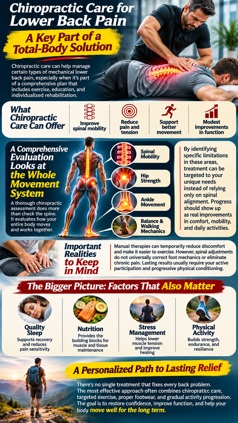
The broader functional perspective also considers sleep quality, nutrition, stress, and physical activity. These factors influence muscular recovery, pain sensitivity, and overall health. Adequate protein supports muscle maintenance, while balanced nutrition contributes to normal tissue function. No specific supplement or detoxification protocol has demonstrated an ability to reliably correct biomechanical back pain.
Rather than searching for one treatment that supposedly fixes every spinal problem, patients benefit from understanding their individual contributors. A successful plan may combine manual care, targeted exercise, footwear adjustments, and gradual activity progression. The objective is restoring confidence and function while reducing the likelihood of recurring limitations. Effective rehabilitation ultimately teaches the body to tolerate movement rather than fear it.
Your At-Home Foot-to-Spine Restoration Plan
Meaningful change begins with a routine that fits into everyday life. Complex exercise programs often fail because people struggle to maintain them consistently. A focused approach can address foot strength, ankle mobility, balance, and hip stability without requiring expensive equipment. The following progression provides a practical foundation for adults with uncomplicated mechanical discomfort.
Begin Each Morning by Restoring Foot Awareness
Before beginning daily activities, spend several minutes gently moving the ankles and toes. Perform slow ankle circles while seated, followed by comfortable big-toe lifts and toe spreading. These movements encourage joint motion and help develop awareness of the feet. They should feel controlled and comfortable rather than aggressive or painful.
Continue with ten short-foot contractions while keeping the toes relaxed against the floor. Hold each contraction for approximately five seconds before releasing. This simple exercise challenges the intrinsic foot muscles without requiring substantial resistance. Gradually progress toward standing practice as coordination improves.
Morning movement does not need to eliminate every sensation of stiffness immediately. Its purpose involves preparing the feet for activity and maintaining consistent muscular engagement. People with significant morning heel pain should avoid aggressive stretching before their symptoms settle. Persistent pain warrants evaluation for conditions such as plantar fasciopathy or inflammatory disorders.
Build Ankle Mobility and Lower-Leg Strength
During the afternoon, dedicate several minutes to ankle mobility and calf strengthening. Perform controlled knee-to-wall movements while maintaining heel contact and comfortable knee alignment. Follow with gentle calf stretches using both straight-knee and bent-knee positions. These variations address different muscular contributions to ankle movement.
Next, perform supported heel raises with slow, controlled lowering. Begin with eight to twelve repetitions and adjust according to your current ability. Strong calf muscles help support walking, balance, and propulsion. Gradual progression improves load tolerance without unnecessarily overwhelming sensitive tissues.
If exercise increases pain substantially or causes lingering symptoms, reduce the intensity or frequency. Recovery depends on matching exercise demands to the body’s present capacity. More repetitions do not automatically produce better results. Consistent, tolerable progression generally provides a stronger foundation for sustainable improvement.
Train Balance and Reconnect the Lower Extremities
Balance training challenges the nervous system to coordinate sensory information and muscular responses. Begin by standing near a sturdy countertop with both feet comfortably positioned. Shift your weight slowly from side to side without excessive trunk movement. Notice how the feet and ankles respond as pressure changes.
When comfortable, practice standing on one leg while keeping a hand near the countertop. Aim for ten to twenty seconds before switching sides. Progress toward longer holds only when balance improves and the exercise remains safe. Individuals with neuropathy, dizziness, or frequent falls should receive professional guidance before attempting unsupported balance challenges.
Balance training may improve postural control and confidence during daily movement. However, better balance does not guarantee relief from chronic lower back pain. Its value lies in improving one component of the broader movement system. Combining balance practice with strength and mobility exercises creates a more complete rehabilitation approach.
Restore Hip Stability and Build Walking Endurance
Finish the routine with glute bridges or another comfortable hip-strengthening exercise. Perform controlled repetitions while avoiding unnecessary spinal strain. Add side-lying hip abduction when appropriate to challenge lateral hip stability. These exercises support the muscular demands of standing, walking, and stair climbing.
Walking provides an opportunity to integrate improvements into everyday movement. Begin with a duration that does not significantly aggravate symptoms, even if that means five minutes. Gradually increase the distance or time as comfort and endurance improve. Consistent exposure helps rebuild physical capacity without demanding immediate perfection.
Avoid consciously forcing an unnatural walking pattern unless a qualified professional identifies a specific reason. Excessive attention to every foot movement can create unnecessary tension and reduce walking comfort. Instead, focus on relaxed movement, appropriate footwear, and gradual increases in activity. Functional improvement should become evident through greater walking tolerance and easier daily activities.
The Long-Term Goal: A Stronger Foundation for a Healthier Spine
Chronic back pain rarely develops through one simple mechanical failure. It may reflect an interaction between tissue sensitivity, physical conditioning, movement habits, previous injuries, and neurological processing. Foot mechanics sometimes contribute to this complicated picture, particularly when symptoms accompany prolonged standing or walking. Recognizing that possibility expands the investigation without reducing every spinal complaint to an arch problem.
The encouraging reality is that the body possesses considerable capacity for adaptation. Muscles strengthen through progressive loading, balance improves with practice, and mobility may increase through appropriate training. These changes can help people perform everyday activities with greater comfort and confidence. Recovery becomes more achievable when rehabilitation addresses relevant limitations instead of chasing perfect posture.
For someone who has spent years concentrating exclusively on the lower back, examining the feet may reveal an important missing piece. An ankle restriction, painful big toe, or weakness during single-leg stance may influence movement farther up the body. Addressing these factors does not guarantee a cure, but it can improve the completeness of treatment. Each meaningful improvement contributes to a stronger, more resilient movement system.
Ultimately, restoring healthier foot mechanics means developing the strength, mobility, coordination, and endurance required for daily life. The spine does not function independently from the structures beneath it. Every step represents a coordinated interaction between the ground, the lower extremities, and the nervous system. By caring for that entire relationship, individuals can pursue better movement and lasting musculoskeletal health.
The foundation beneath your body deserves the same attention as the structures above it. When the feet function comfortably and the muscles throughout the kinetic chain work effectively, movement often becomes easier. Rather than endlessly treating symptoms in isolation, investigate the mechanical relationships that influence them. A healthier back may begin with understanding how your body meets the ground.