
The Foundation That Could Be Fueling Your Back Pain
A house can have beautiful walls, a flawless roof, and expensive windows, yet still develop cracks throughout its structure. When the foundation shifts, everything resting above it must adapt to the changing distribution of weight. Contractors understand that repairing visible cracks without investigating the foundation often leads to repeated damage. The human spine faces a similar mechanical challenge when the pelvis cannot effectively support and coordinate movement.
Consider a patient who has battled chronic lower back pain for years. Every morning begins with stiffness, and prolonged sitting makes standing uncomfortable. Simple activities, including gardening or lifting groceries, trigger recurring muscle spasms. Massage provides temporary relief, while pain medication quiets the discomfort without explaining its origin. Despite numerous treatments, the underlying movement problem may remain unidentified.
The missing connection sometimes lies within the pelvis, the powerful structural bridge connecting the spine to the legs. Its position influences spinal curvature, muscular demands, balance, and the distribution of mechanical forces. When pelvic movement becomes restricted or poorly coordinated, surrounding tissues may compensate during ordinary activities. Over time, these compensations can contribute to fatigue, discomfort, and reduced movement tolerance.
However, a tilted pelvis does not automatically mean something is damaged or out of place. Healthy people demonstrate considerable variation in pelvic alignment, and many experience no pain whatsoever. The real concern arises when pelvic mechanics interact with weakness, restricted mobility, previous injuries, or excessive physical demands. Understanding these relationships offers a more meaningful starting point for investigating persistent back pain.
Understanding Pelvic Tilt: The Structural Connection Between Your Hips and Spine
The pelvis forms the foundation of the spinal column and serves as a central platform for human movement. Two hip bones connect with the sacrum, creating a strong ring that transfers forces between the trunk and legs. Above this structure sit five lumbar vertebrae, which support movement while protecting important neurological tissues. Below it, the hip joints provide the mobility required for walking, bending, climbing, and lifting.
Surrounding the pelvis are powerful muscles responsible for stability, posture, and coordinated movement. The gluteal muscles generate hip extension and help control the pelvis during walking. Abdominal muscles contribute to trunk stability, while deep spinal muscles assist with movement and segmental control. Hip flexors, including the iliopsoas, connect the lower body with structures surrounding the lumbar spine.
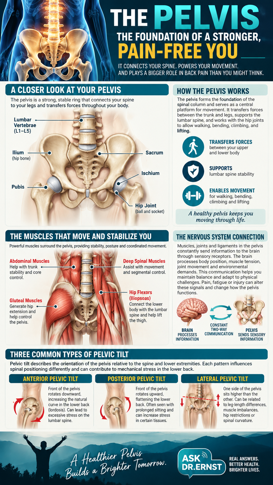
These muscles constantly exchange information with the nervous system through sensory receptors embedded throughout the musculoskeletal system. The brain processes information about body position, muscular tension, joint movement, and environmental demands. This communication allows the body to maintain balance while adapting to different physical challenges. Pain, fatigue, or injury can alter these movement strategies and change how the pelvis functions.
Pelvic tilt describes the orientation of the pelvis relative to the spine and lower extremities. Three common patterns include anterior pelvic tilt, posterior pelvic tilt, and lateral pelvic tilt. Each pattern influences spinal positioning differently and may become relevant during a clinical assessment. Understanding their characteristics helps reveal how mechanical stress can develop throughout the lower back.
Anterior Pelvic Tilt: When the Lower Back Works Overtime
Anterior pelvic tilt occurs when the front of the pelvis rotates downward while the back rotates upward. This position often increases the inward curvature of the lumbar spine, known as lumbar lordosis. A pronounced arch may become visible around the lower back, sometimes accompanied by a forward-projecting abdomen. Although this posture can appear unusual, its presence alone does not establish a medical problem.
Prolonged sitting, limited hip extension, and habitual movement patterns may contribute to anterior pelvic positioning in certain individuals. Reduced hip mobility can encourage additional lumbar movement during walking, standing, or reaching overhead. When surrounding muscles lack sufficient endurance, maintaining these positions may become increasingly uncomfortable. Repetitive extension-sensitive activities can then aggravate vulnerable joints or other pain-sensitive tissues.
The iliopsoas deserves attention because its psoas major component attaches directly to the lumbar vertebrae. This anatomical relationship allows the muscle to influence hip movement and spinal mechanics. Restricted hip extension may encourage some individuals to compensate through the lower back. Nevertheless, tight hip flexors do not automatically cause anterior pelvic tilt or chronic pain.
The important question concerns how the spine responds to physical demands rather than how dramatically it curves. A patient who develops pain during prolonged standing may demonstrate reduced hip mobility or limited trunk endurance. Another individual with identical pelvic alignment may remain completely comfortable during demanding exercise. Functional testing helps distinguish a meaningful mechanical problem from a harmless anatomical variation.
Posterior Pelvic Tilt: When the Spine Loses Its Natural Curve
Posterior pelvic tilt occurs when the front of the pelvis rotates upward and the lower back becomes flatter. This position commonly appears during slouched sitting, abdominal bracing, and certain exercise movements. Some individuals naturally stand with reduced lumbar lordosis, while others adopt this position to avoid discomfort. The mechanical consequences depend on the person’s anatomy, symptoms, and physical activities.
Prolonged spinal flexion may increase discomfort in individuals sensitive to bending or sustained sitting. Lumbar discs, ligaments, muscles, and other tissues experience changing loads as spinal position changes. Repeated exposure can become problematic when those loads exceed a person’s current tolerance. However, a flatter lumbar spine does not automatically indicate degeneration, weakness, or future injury.
Individuals with posterior pelvic tilt may also demonstrate limited hip movement or altered hamstring and abdominal muscle coordination. These relationships vary considerably and require direct examination rather than assumptions based on appearance. Excessive emphasis on maintaining one ideal spinal curve can create unnecessary fear surrounding normal movement. Healthy backs need the ability to flex, extend, rotate, and adapt comfortably.
Lateral Pelvic Tilt: The Hidden Imbalance Between Your Hips
Lateral pelvic tilt occurs when one side of the pelvis appears higher than the other. This asymmetry may develop through differences in leg length, hip mobility, muscular control, or spinal curvature. Previous injuries and habitual weight shifting can also influence how the pelvis appears during standing. In some individuals, scoliosis contributes to a visible difference between the two sides.
During walking, the gluteus medius helps stabilize the pelvis as body weight transfers onto one leg. Reduced hip abductor strength or impaired coordination may allow excessive pelvic dropping during this movement. The trunk may compensate by leaning sideways, changing the forces acting across the lumbar spine. Repeated compensation can become uncomfortable when strength and endurance cannot meet physical demands.
Nevertheless, small differences between the left and right sides occur naturally throughout the human body. A slightly elevated hip does not prove that the pelvis requires realignment. Even measurable leg-length differences may produce no symptoms in otherwise healthy individuals. Clinical significance depends on the magnitude of the difference, associated symptoms, and functional limitations.
The Sacroiliac Joint: A Hidden Contributor to Pelvic and Lower Back Pain
Deep within the posterior pelvis, the sacrum connects to both hip bones through the sacroiliac joints. Powerful ligaments stabilize these joints while permitting small movements that support force transfer during daily activities. Although their movement is limited, the sacroiliac joints withstand considerable mechanical demands during walking, climbing, and lifting. Irritation within this region can produce discomfort around the lower back, buttocks, and sometimes the thighs.
Sacroiliac pain often resembles discomfort originating from lumbar discs, spinal joints, or surrounding muscles. Patients may experience pain when standing from a chair, climbing stairs, or shifting weight onto one leg. Pregnancy, previous injuries, inflammatory disorders, and repetitive loading can contribute to sacroiliac symptoms. Consequently, an accurate diagnosis requires more than observing whether one hip appears higher.
Some patients hear that their sacrum has slipped out of place or their pelvis requires repeated repositioning. Yet normal sacroiliac joints possess substantial ligamentous stability and rarely undergo true displacement without major trauma. Pain usually reflects a more complex interaction involving sensitive tissues, mechanical loading, and neurological responses. Appropriate examination helps determine whether the sacroiliac joint actually contributes to the patient’s symptoms.
The Nervous System Connection: When Mechanical Stress Becomes Chronic Pain
The nervous system continuously monitors movement, muscular tension, and changing forces throughout the pelvis and spine. Sensory receptors transmit information that helps the brain coordinate balance, stability, and protective responses. Following injury, pain can change muscle activation and encourage guarding around sensitive areas. These adaptations may initially protect the body but can become counterproductive when they persist unnecessarily.
Chronic discomfort sometimes develops alongside increased nervous system sensitivity, making ordinary movement feel unusually threatening or painful. Muscles may remain guarded, movement confidence can decline, and physical activity often decreases. Reduced activity gradually weakens muscular endurance, leaving the body less prepared for everyday physical demands. This cycle can perpetuate disability even after the original injury has improved.
The consequences extend beyond the lower back, influencing walking tolerance, sleep quality, work performance, and overall independence. Persistent discomfort can discourage exercise, contributing to declining cardiovascular fitness and reduced muscular conditioning. Sleep disruption may further amplify pain sensitivity and interfere with recovery. Without addressing these interconnected factors, temporary symptom relief may fail to produce lasting functional improvement.
The Real Root Cause: A Pelvis That Cannot Adapt to Life’s Demands
The greatest misconception surrounding pelvic tilt is that one abnormal-looking posture explains every case of chronic back pain. Research demonstrates that people with and without lower back pain frequently share similar resting pelvic positions. More meaningful differences sometimes emerge through movement speed, spinal mobility, proprioception, and functional performance. Therefore, identifying the underlying problem requires investigating how the entire musculoskeletal system responds to physical demands.
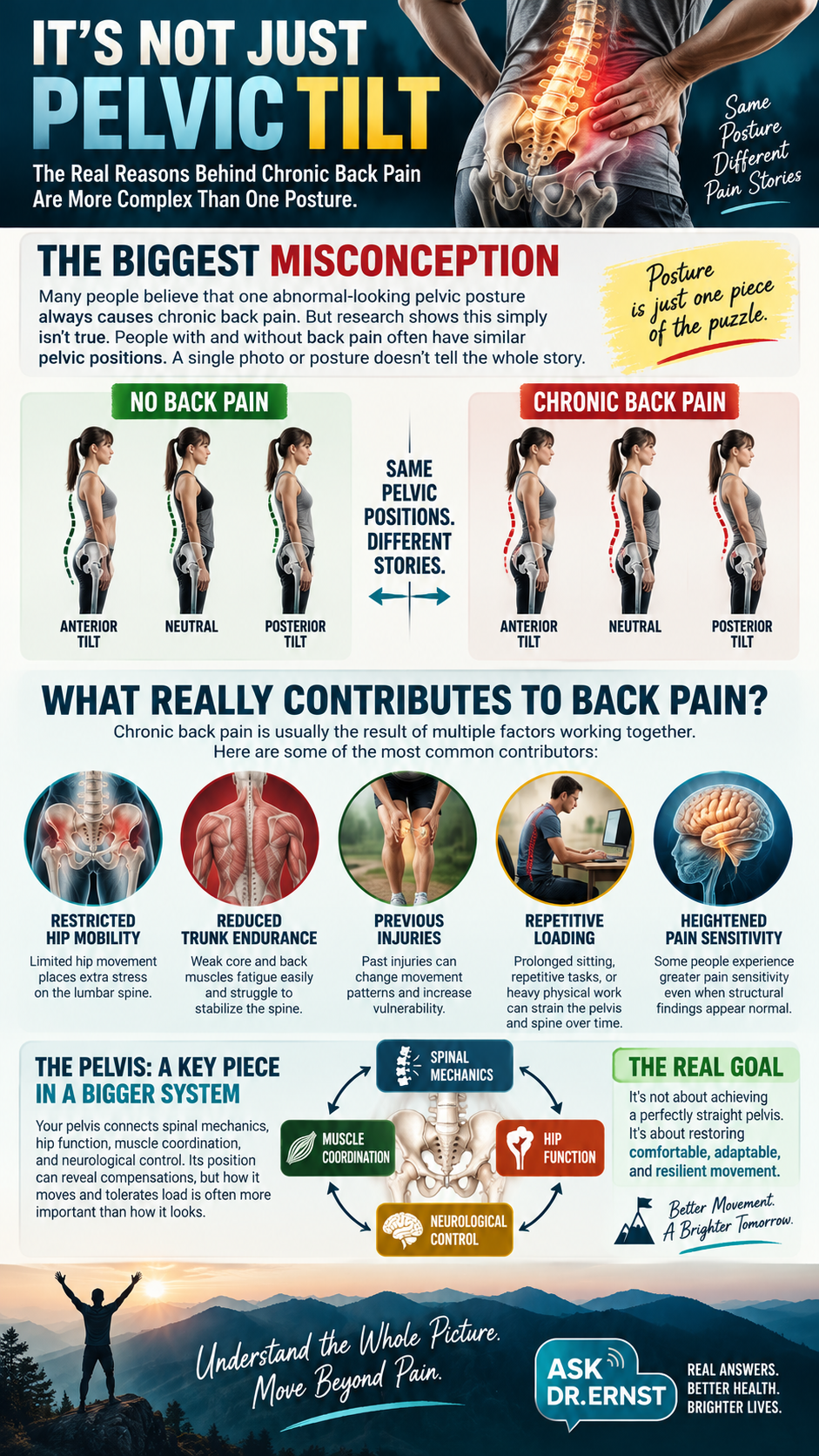
For some patients, restricted hip mobility creates excessive demands on the lumbar spine during everyday movement. Others struggle with reduced trunk endurance, previous injuries, poor conditioning, or repetitive occupational loading. Certain individuals experience heightened pain sensitivity despite relatively normal structural findings. Each situation requires a different explanation and an individualized recovery strategy.
The pelvis offers valuable clues because it connects spinal mechanics, hip function, muscular coordination, and neurological control. Its position may reveal compensations, but its ability to move and tolerate load often provides more useful information. The ultimate objective is not achieving a perfectly straight pelvis; it is restoring comfortable, adaptable, and resilient movement. Understanding that distinction opens the door to addressing chronic back pain beyond temporary symptom management.
The Path to Healing Begins With Understanding the Cause
Chronic back pain rarely develops from one isolated problem. Instead, years of repetitive movements, restricted mobility, muscular imbalances, and previous injuries can gradually alter how the body functions. The pelvis often sits at the center of these mechanical relationships, connecting spinal movement with lower-extremity stability. When this system loses its ability to adapt, ordinary activities can become exhausting, uncomfortable, and increasingly difficult.
Many patients spend years chasing temporary relief without understanding the physical limitations contributing to their discomfort. Massage may relax tense muscles, while medication can reduce pain long enough to resume daily activities. However, neither approach necessarily improves limited hip mobility, reduced muscular endurance, or inefficient movement strategies. Lasting improvement often requires identifying these limitations and progressively restoring the body’s capacity to tolerate physical demands.
The most effective approach begins with a comprehensive examination rather than assuming every tilted pelvis needs correction. Spinal mobility, hip function, neurological findings, muscular strength, and movement tolerance all deserve careful evaluation. An individual’s symptoms, medical history, and daily activities help determine which findings actually matter. This personalized approach provides a stronger foundation for meaningful recovery than pursuing an arbitrary standard of perfect posture.
Chiropractic Care: Restoring Movement Throughout the Spine and Pelvis
Chiropractic care offers one conservative approach to evaluating and managing mechanical lower back pain. A thorough examination can identify painful spinal movements, restricted joint mobility, and functional limitations involving the pelvis. Chiropractors may also assess gait, muscular endurance, hip movement, and neurological function. These findings help determine whether spinal manipulation, exercise, or additional medical evaluation may be appropriate.
Spinal manipulation applies a controlled mechanical force to selected joints, potentially influencing pain sensitivity and movement tolerance. Clinical research suggests that manipulation can provide modest improvements in pain and function for certain patients. However, treatment does not reliably reposition pelvic bones into a permanently corrected anatomical alignment. Its greatest value may emerge when clinicians combine appropriate manual therapy with progressive rehabilitation and patient education.
Consider someone whose lower back becomes painful after standing for extended periods. Examination might reveal limited hip extension, reduced abdominal endurance, and discomfort during repeated lumbar extension. Manual therapy could temporarily improve movement comfort, creating an opportunity to begin targeted strengthening exercises. Continued improvement would depend largely on developing physical capacity and addressing the activities that repeatedly aggravate symptoms.
An important distinction separates improving spinal function from claiming that every spinal restriction causes widespread organ dysfunction. The nervous system coordinates movement, sensation, and numerous physiological processes, but pelvic tilt does not establish nerve interference. Effective chiropractic care should emphasize measurable improvements in pain, mobility, strength, and everyday function. These outcomes provide patients with meaningful evidence that their recovery is progressing.
The Hip Flexor Problem: Restoring Mobility Without Forcing Alignment
The hip flexors influence pelvic movement because several muscles cross the front of the hip joint. The iliopsoas helps lift the thigh, while the rectus femoris contributes to both hip flexion and knee extension. Prolonged sitting may reduce opportunities to move these muscles through their available range. Some individuals consequently develop hip extension limitations that influence walking, standing, or exercise mechanics.
Restricted hip extension can encourage compensatory movement through the lumbar spine during certain activities. For someone with extension-sensitive back pain, this strategy may increase discomfort during prolonged standing or walking. However, tight hip flexors do not automatically explain anterior pelvic tilt. Treatment should target a demonstrated mobility restriction rather than an assumption based on posture.
Gentle stretching can improve flexibility when clinicians identify meaningful restrictions. Strengthening through comfortable ranges may also help the hip flexors tolerate physical demands more effectively. Excessive stretching, particularly when it increases back discomfort, can aggravate symptoms rather than improve them. The goal involves developing useful mobility and muscular control, not forcing the pelvis into a predetermined position.
Gluteal Strength: Rebuilding the Support System Beneath Your Spine
The gluteal muscles provide essential support during walking, climbing stairs, lifting, and standing on one leg. Gluteus maximus generates powerful hip extension, while gluteus medius helps control pelvic movement during weight transfer. When these muscles lack sufficient strength or endurance, other structures may compensate during demanding activities. This compensation can contribute to discomfort in individuals whose tissues cannot tolerate the additional loading.
Weak gluteal muscles do not necessarily cause pelvic tilt, and visible asymmetry does not prove muscular weakness. Nevertheless, targeted strengthening can improve physical capacity and support rehabilitation for many people with chronic back pain. Exercises should progress according to symptoms, movement quality, and individual fitness levels. Developing stronger hips often helps patients perform everyday activities with greater confidence and less fatigue.
Bridges, controlled squats, step-ups, and resistance-band exercises provide several ways to challenge the gluteal muscles. Initially, simple movements may offer sufficient resistance for someone recovering from prolonged inactivity. Gradually increasing repetitions, resistance, and movement complexity encourages muscular adaptation. Consistency matters more than performing aggressive workouts that repeatedly trigger painful flare-ups.
The Functional Medicine Connection: Supporting Recovery From Within
Musculoskeletal recovery depends on more than the position of bones and joints. Sleep quality, nutrition, physical conditioning, and metabolic health influence muscular performance and pain sensitivity. Persistent sleep disruption can intensify discomfort, while inadequate nutrition may compromise the body’s ability to maintain muscle. Addressing these factors creates a healthier environment for rehabilitation.
Protein supports muscular repair and adaptation during progressive resistance training. Nutrient-dense foods provide essential vitamins, minerals, and energy needed for normal tissue function. Omega-3-rich fish, vegetables, olive oil, and other minimally processed foods can contribute to a balanced dietary pattern. However, no particular supplement or detoxification protocol has demonstrated an ability to permanently correct pelvic tilt.
Stress management also deserves consideration because persistent stress can disrupt sleep and influence pain processing. Breathing exercises, regular movement, and consistent sleep schedules may help reduce the burden of chronic discomfort. These practices complement physical rehabilitation rather than replacing it. A comprehensive strategy addresses the whole person while remaining focused on measurable improvements in physical function.
Practical At-Home Exercises to Improve Pelvic Tilt and Lower Back Pain
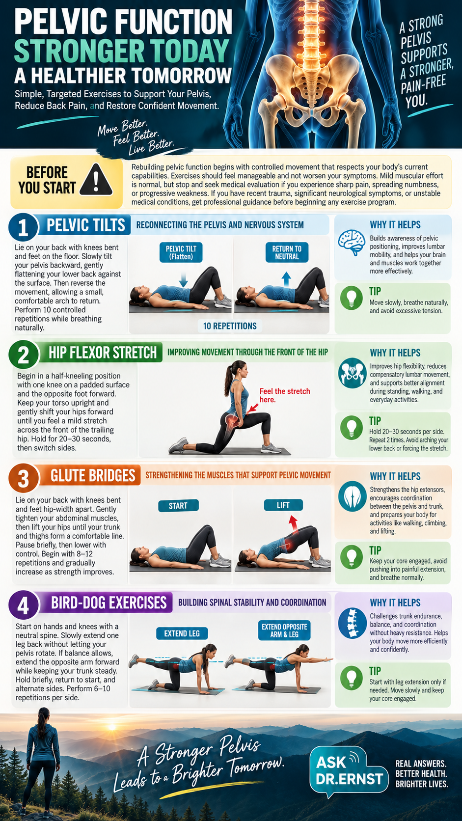
Rebuilding pelvic function begins with controlled movement that respects the body’s current capabilities. Exercises should feel manageable and produce no substantial worsening of symptoms. Mild muscular effort is expected, but sharp pain, spreading numbness, or progressive weakness warrants stopping and seeking evaluation. Individuals with recent trauma, significant neurological symptoms, or unstable medical conditions need professional guidance before beginning rehabilitation.
Pelvic Tilts: Reconnecting the Pelvis and Nervous System
Lie comfortably on your back with both knees bent and feet resting on the floor. Slowly rotate the pelvis backward, gently flattening the lower back against the surface. Reverse the movement by allowing a small, comfortable arch to return. Perform ten controlled repetitions while breathing naturally and avoiding excessive muscular tension.
This movement helps develop awareness of pelvic positioning and comfortable lumbar mobility. Repeated practice may improve coordination and reduce fear surrounding spinal movement. The exercise does not physically reset displaced pelvic bones or guarantee lasting postural correction. Its purpose is to restore comfortable movement options that support everyday activities.
Hip Flexor Stretching: Improving Movement Through the Front of the Hip
Begin in a half-kneeling position with one knee supported on a padded surface. Place the opposite foot forward and maintain a comfortable, upright trunk position. Gently shift your hips forward until you notice mild tension across the front of the trailing hip. Hold for twenty to thirty seconds before changing sides.
Avoid arching the lower back excessively or forcing the hips beyond a comfortable range. Repeat the stretch twice per side when appropriate, particularly after extended sitting. Improved hip flexibility may reduce compensatory lumbar movement in individuals with restricted hip extension. Stretching should remain one component of rehabilitation rather than the entire treatment strategy.
Glute Bridges: Strengthening the Muscles That Support Pelvic Movement
Lie on your back with knees bent and feet positioned approximately hip-width apart. Tighten your abdominal muscles gently, then lift your hips until your trunk and thighs form a comfortable line. Pause briefly before lowering your pelvis with control. Begin with eight to twelve repetitions and gradually increase as strength improves.
Glute bridges strengthen the hip extensors while encouraging coordinated movement between the pelvis and trunk. Avoid pushing into painful lumbar extension or holding your breath throughout the exercise. As tolerance improves, resistance bands or additional repetitions can increase the challenge. Progressive strengthening helps prepare the hips for walking, climbing, and lifting.
Bird-Dog Exercises: Building Spinal Stability and Coordination
Position yourself on hands and knees while maintaining a comfortable spinal posture. Slowly extend one leg backward without allowing excessive rotation through the pelvis. If balance permits, extend the opposite arm forward while keeping the trunk steady. Hold briefly, return to the starting position, and alternate sides.
Perform approximately six to ten repetitions per side with deliberate control. This exercise challenges trunk endurance, balance, and coordination without requiring heavy external resistance. Begin with leg movements alone when simultaneous arm extension feels difficult. Gradually increasing control and endurance can improve confidence during more demanding activities.
Everyday Habits That Protect Your Back From Recurring Pain
Exercise sessions represent only one part of the recovery process. Daily movement habits influence how frequently the spine and pelvis experience sustained or repetitive loading. Remaining seated for several hours without changing position may aggravate symptoms in some individuals. Brief walking breaks and regular position changes can help maintain comfort throughout the day.
Walking provides an accessible way to improve conditioning while gradually increasing tolerance for movement. Begin with manageable distances and progress according to symptoms and recovery. Avoid treating every uncomfortable sensation as evidence of additional structural damage. Appropriate activity often supports recovery better than prolonged bed rest.
During lifting, keep objects reasonably close to the body and use a movement strategy that feels controlled. Allow the hips, knees, and spine to share the demands of the task. No single lifting posture guarantees protection from injury, and normal spinal movement should not inspire fear. Gradual strength development and sensible load progression matter more than maintaining rigid posture.
When Pelvic Tilt Signals Something More Serious
Not every case of lower back pain originates from mechanical dysfunction. Persistent symptoms accompanied by fever, unexplained weight loss, or a history of cancer require medical assessment. New bowel or bladder dysfunction, saddle numbness, or progressive leg weakness may indicate neurological emergencies. These symptoms demand urgent evaluation rather than stretching, manipulation, or home exercises.
Inflammatory spinal conditions can also produce persistent discomfort, particularly when morning stiffness improves with activity. Hip arthritis, lumbar nerve compression, and other disorders may resemble pelvic-related mechanical pain. Appropriate examination helps distinguish these conditions and guides further testing when necessary. Identifying the correct diagnosis protects patients from pursuing ineffective treatment for the wrong problem.
Healing the Foundation: Building a Back That Can Move Without Fear
The pelvis tells an important story about the relationship between spinal mechanics, muscular function, and everyday physical demands. Its position may reveal movement adaptations, but appearance alone cannot identify the cause of chronic pain. Real progress comes from understanding how the hips, spine, muscles, and nervous system work together. Rehabilitation should improve their collective ability to handle the activities that matter most.
Chiropractic care, targeted strengthening, improved mobility, and supportive lifestyle changes can contribute to meaningful recovery. No single intervention guarantees a permanently corrected pelvis or complete elimination of back pain. Yet consistent, individualized care can improve physical function, reduce disability, and restore confidence in movement. The objective is not a perfectly aligned skeleton, but a resilient body capable of adapting comfortably to life.
Your pelvis does not need to look perfect to support a healthy spine. It needs sufficient mobility, strength, coordination, and tolerance for the demands you place upon it. When rehabilitation addresses these qualities, chronic back pain no longer needs to dictate every movement or decision. That is the foundation of sustainable recovery and a healthier, more capable future.