
The Question Few Women Ever Hear
Sarah had barely finished buttoning her blouse when the technician smiled and said, “We’ll see you again next year.” The appointment lasted less than thirty minutes, yet its meaning stretched far beyond that brief visit. Like millions of women before her, she walked out believing she had done exactly what every responsible woman should do. She never questioned the machine, the process, or the science behind it. She trusted decades of public health campaigns, physician recommendations, and television advertisements that repeated one simple message: annual mammograms save lives. Few people challenged that narrative because it sounded logical, reassuring, and rooted in modern medicine. Yet beneath that confidence lies a more complicated story, one that deserves careful examination rather than unquestioned acceptance.
Breast cancer remains one of the most feared diseases among women because its diagnosis changes lives in an instant. That fear has driven enormous investments into screening technologies designed to find tumors as early as possible. Physicians rightly understand that cancers discovered at an earlier stage often require less aggressive treatment and generally produce better outcomes. Early detection has therefore become the cornerstone of breast cancer prevention campaigns around the world. Unfortunately, the phrase “early detection” often distracts from another important question. What if the body signals trouble years before a tumor becomes visible? Even more importantly, what if those early biological changes offer an opportunity to intervene before structural disease develops?
Modern medicine has traditionally focused on finding disease after it becomes visible. Functional medicine approaches the problem from a different direction by asking why disease develops in the first place. Instead of waiting for tissue to become abnormal enough to appear on an image, functional clinicians search for disturbances in physiology that precede anatomical damage. That distinction represents far more than a philosophical difference. It reflects two fundamentally different ways of thinking about health. One waits for disease to declare itself. The other attempts to recognize dysfunction while restoration remains possible.
This difference explains why mammography and thermography often generate passionate debate. Supporters of mammography emphasize decades of research showing improved detection of many breast cancers. Advocates of thermography argue that physiology changes long before anatomy does, making functional imaging an attractive addition to preventive care. Unfortunately, discussions surrounding these technologies often become emotional rather than scientific. One side sometimes portrays mammography as flawless, while the other occasionally describes thermography as a complete replacement. Neither position accurately reflects the complexity of breast biology or the current scientific literature.
Understanding that complexity begins by recognizing a simple truth. Breast cancer does not suddenly appear on the day a radiologist identifies a suspicious shadow. The disease develops through years of microscopic biological change that quietly reshape the tissue long before imaging reveals a recognizable mass. Appreciating that timeline changes the entire conversation about screening because it shifts attention away from finding established disease and toward understanding how disease develops.
Cancer Begins as a Biological Process, Not an Imaging Finding
Every healthy cell within the breast follows an intricate set of biological instructions encoded within its DNA. Those instructions regulate growth, repair damaged tissue, coordinate immune responses, and determine when aging cells should naturally die. Healthy tissue maintains remarkable balance because billions of cells communicate continuously through hormones, growth factors, neurotransmitters, immune signals, and electrical impulses. That communication allows the breast to adapt throughout life, particularly during puberty, pregnancy, breastfeeding, and menopause.
Problems arise when those regulatory systems begin breaking down. Chronic inflammation, oxidative stress, hormonal imbalance, environmental toxins, poor metabolic health, and accumulated genetic damage gradually interfere with normal cellular communication. Individual cells may begin dividing more rapidly than they should. Others lose their ability to repair damaged DNA efficiently. Some escape the natural process of programmed cell death, allowing abnormal cells to survive when healthy cells would normally disappear. None of these changes create an immediate tumor. Instead, they establish the biological conditions that allow cancer to emerge over time.
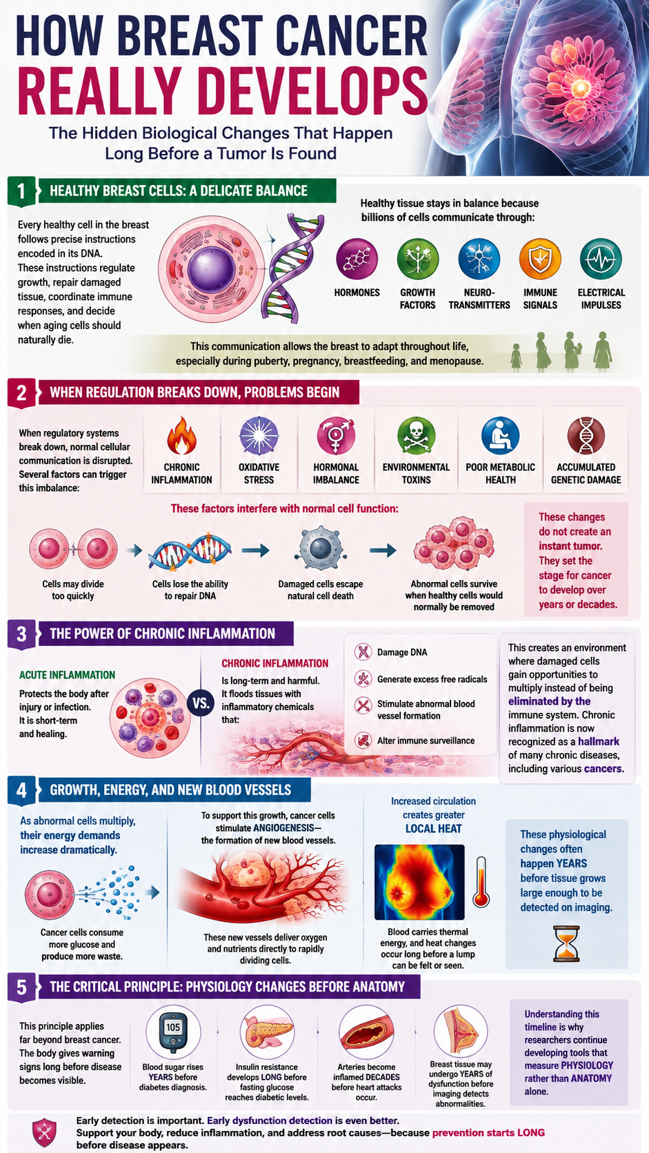
Inflammation deserves particular attention because it influences nearly every stage of cancer development. Acute inflammation protects the body after injury or infection. Chronic inflammation behaves very differently. It floods tissues with inflammatory chemicals that damage DNA, generate excessive free radicals, stimulate abnormal blood vessel formation, and alter immune surveillance. These changes create an environment where damaged cells gain opportunities to multiply instead of being removed by the immune system. Scientists increasingly recognize chronic inflammation as one of the hallmarks of many chronic diseases, including numerous forms of cancer.
As abnormal cells continue multiplying, their energy demands increase dramatically. Cancer cells consume substantially more glucose than healthy cells while producing excessive metabolic waste. To support this accelerated growth, they stimulate angiogenesis, the formation of new blood vessels that deliver oxygen and nutrients directly to rapidly dividing tissue. Increased circulation naturally produces greater local heat because blood transports thermal energy throughout the body. These physiological changes often occur long before the tissue grows large enough to create a detectable lump.
This sequence illustrates an important principle that extends far beyond breast cancer. Physiology changes before anatomy changes. Blood sugar often becomes abnormal years before diabetes receives a diagnosis. Insulin resistance develops long before fasting glucose reaches diabetic levels. Arteries become inflamed decades before heart attacks occur. Likewise, breast tissue may undergo years of biological dysfunction before conventional imaging identifies structural abnormalities. Recognizing this timeline explains why many researchers continue exploring technologies capable of measuring physiology rather than anatomy alone.
The Rise of Mammography and Its Lasting Influence
The story of mammography reflects one of medicine’s most significant technological achievements. Early physicians struggled to identify breast tumors before they became large enough to feel during examination. During the twentieth century, advances in X-ray technology created an opportunity to visualize internal breast structures without surgery. Early machines produced grainy images while exposing women to considerably higher radiation doses than today’s equipment. Engineers steadily refined detector technology, image processing, and radiation efficiency, transforming mammography into a sophisticated diagnostic tool capable of identifying remarkably small structural changes.
Large population studies performed throughout the latter half of the twentieth century helped establish mammography as the dominant screening method. Researchers demonstrated that many breast cancers could be detected before producing symptoms, allowing treatment to begin at earlier stages. Public health officials embraced those findings because earlier intervention frequently improved outcomes across large populations. Governments invested heavily in awareness campaigns, physicians incorporated screening into preventive care, and insurance companies expanded coverage for routine examinations. Mammography gradually became more than a diagnostic tool. It became a cultural expectation associated with responsible healthcare.
Technological improvements continued accelerating during the digital era. Film systems gave way to digital detectors capable of producing clearer images with lower radiation exposure. Three-dimensional tomosynthesis reduced tissue overlap by reconstructing thin image slices from multiple angles. Artificial intelligence now assists radiologists by highlighting subtle abnormalities that deserve closer evaluation. These innovations represent genuine progress that improved diagnostic performance while reducing several limitations associated with earlier generations of equipment.
Despite those advances, mammography still shares one defining characteristic with every anatomical imaging technology. It identifies changes after tissue structure becomes abnormal. Calcifications, masses, architectural distortion, and density differences become visible because disease has already altered the physical organization of the breast. Mammography excels at recognizing these structural findings, yet it cannot directly measure inflammation, metabolism, immune activity, oxidative stress, or cellular energy production. Those physiological events occur beyond the reach of conventional X-ray imaging, even though they often precede visible structural disease.
Understanding this distinction does not diminish the value of mammography. Instead, it clarifies what the technology was designed to accomplish. Mammography answers the question, “Has the tissue changed structurally?” It does not answer the equally important question, “Is the biology of this tissue beginning to change before anatomy follows?” That second question has inspired growing interest in complementary technologies capable of evaluating breast physiology from a different perspective.
Radiation: Small Doses That Deserve Honest Discussion
Few words in medicine provoke stronger emotional reactions than radiation. Images of nuclear accidents and cancer treatments often dominate public imagination, making every discussion about medical imaging emotionally charged. Scientific conversations require greater precision because not all radiation exposures carry equal biological significance. Mammography uses low-dose ionizing radiation to generate detailed images of breast tissue. The dose remains substantially lower than many computed tomography examinations, yet it still belongs to the same category of radiation capable of altering DNA.
Ionizing radiation carries enough energy to remove electrons from atoms, producing highly reactive molecules known as free radicals. These unstable molecules can damage DNA, proteins, and cellular membranes when they accumulate faster than antioxidant systems can neutralize them. Human biology fortunately includes sophisticated repair mechanisms that identify and correct most DNA injuries before permanent mutations develop. Every day, normal metabolism produces thousands of microscopic DNA lesions within every cell. Specialized repair enzymes continuously correct those errors with remarkable efficiency.
Difficulties emerge when damage accumulates faster than repair systems can respond. Age naturally reduces repair efficiency. Chronic inflammation increases oxidative stress. Nutritional deficiencies weaken antioxidant defenses. Certain inherited genetic mutations impair DNA repair pathways. These factors do not guarantee cancer development, yet they influence how effectively the body responds to repeated cellular injury from both internal metabolism and external environmental exposures.
Modern digital mammography exposes women to relatively small amounts of radiation during each examination. Supporters correctly emphasize that the dose remains low compared with many other diagnostic procedures. However, radiation biology does not operate according to an all-or-nothing principle. Every exposure contributes incrementally to an individual’s cumulative lifetime radiation burden. Natural background radiation, dental imaging, CT scans, fluoroscopy, airport security systems, occupational exposure, and mammography collectively shape that lifelong profile. Scientists continue studying how repeated low-dose exposures influence long-term cancer risk across diverse populations.
Research has generated particular interest regarding women carrying BRCA1 and BRCA2 mutations because those genes help repair damaged DNA. Some investigators have proposed that impaired repair capacity could theoretically increase sensitivity to ionizing radiation. Several studies have suggested greater vulnerability among genetically susceptible individuals, while others have reported smaller associations than initially expected. Current medical guidelines continue recommending appropriate imaging for high-risk women because available evidence indicates that the benefits of surveillance generally outweigh the estimated radiation risks. Nevertheless, ongoing investigation reflects an important reality. The scientific community continues refining its understanding of cumulative low-dose radiation rather than declaring every question permanently settled.
Balanced discussion requires avoiding both extremes. Describing mammography as harmless ignores the well-established biology of ionizing radiation. Portraying routine screening as highly dangerous ignores decades of evidence demonstrating its clinical usefulness. Honest science rarely supports absolute conclusions. Instead, it encourages informed decision-making based upon individual risk factors, family history, breast density, age, genetics, and overall health. Understanding those variables allows women to participate actively in decisions affecting their long-term breast health rather than relying solely upon generalized recommendations.
Thermography Looks at Function Before Structure
Imagine standing outside a home on a cold winter evening. Every wall appears solid, every window seems intact, and the roof looks perfectly sound. An infrared camera, however, tells a different story. Bright patches of escaping heat reveal hidden gaps behind the walls that the human eye cannot see. The structure appears normal, yet the function has already begun to fail. Thermography applies that same principle to the human body by measuring patterns of infrared heat rather than photographing internal anatomy.
Every living cell produces heat while generating energy. Healthy tissue maintains predictable temperature patterns because circulation, nerve activity, and metabolism remain balanced. When inflammation develops, blood vessels expand to deliver immune cells and nutrients into the affected area. Metabolism increases as tissues demand more energy for repair. These physiological changes create localized heat that sophisticated infrared cameras can detect without touching the body. Instead of looking for a mass, thermography evaluates biological activity occurring beneath the skin.
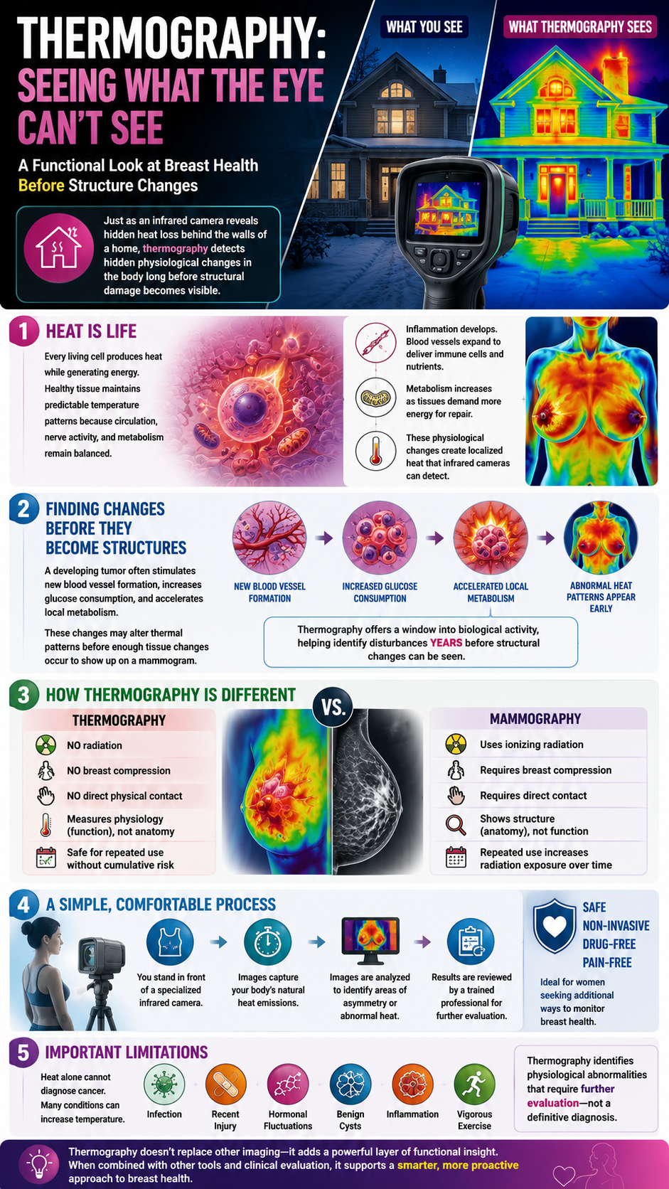
Supporters of thermography believe this functional approach offers an opportunity to identify physiological disturbances before structural damage becomes obvious. A developing tumor often stimulates new blood vessel formation, increases glucose consumption, and accelerates local metabolism. Those changes may alter thermal patterns before enough tissue changes occur to produce an abnormal mammogram. This concept explains why thermography continues attracting interest among clinicians who emphasize prevention rather than disease management.
Thermography also differs from mammography in several practical ways. The examination does not compress breast tissue, expose patients to ionizing radiation, or require direct physical contact. The procedure typically involves standing in front of a specialized infrared camera while images record the body’s natural heat emissions. Because the technology measures physiology rather than anatomy, repeated examinations do not increase radiation exposure. That feature appeals to women seeking additional ways to monitor breast health without cumulative imaging dose.
Despite these advantages, thermography also has important limitations. Heat alone cannot diagnose cancer because many biological processes increase local temperature. Infection, recent injury, hormonal fluctuations, benign cysts, inflammation, and even vigorous exercise may influence thermal patterns. Thermography therefore identifies physiological abnormalities that require further evaluation rather than providing a definitive diagnosis. Understanding that distinction prevents unrealistic expectations while highlighting the technology’s potential role within a broader breast health strategy.
What the Scientific Literature Really Shows
Few medical topics generate stronger opinions than breast cancer screening. Unfortunately, opinions often overshadow careful analysis of scientific evidence. Some advertisements describe thermography as a complete replacement for mammography, while others dismiss it entirely. Current research supports neither extreme. Instead, the literature paints a far more nuanced picture.
Numerous studies have demonstrated that thermography detects abnormal physiological activity associated with increased blood flow, inflammation, and metabolic change. Several investigations have reported that persistent abnormal thermal patterns correlate with an elevated future risk of developing breast cancer. These findings suggest thermography may identify biological disturbances deserving closer observation before structural abnormalities appear. Researchers continue exploring whether combining functional and anatomical imaging could improve overall screening strategies.
At the same time, studies have also demonstrated that thermography lacks sufficient sensitivity and specificity to function as a standalone screening test for breast cancer in the general population. Some cancers produce little measurable thermal change, particularly small tumors located deep within the breast. Conversely, many noncancerous conditions generate abnormal heat patterns that may lead to unnecessary concern if interpreted without additional clinical information. These limitations explain why major medical organizations currently recommend against replacing mammography with thermography alone.
This distinction often becomes misunderstood. Rejecting thermography as a replacement does not imply the technology lacks value. Electrocardiograms do not replace echocardiograms. Blood tests do not replace MRI scans. Ultrasound does not replace colonoscopy. Different technologies answer different biological questions. Thermography evaluates function. Mammography evaluates structure. Combining multiple sources of information often produces a more complete understanding than relying upon any single test in isolation.
Scientific progress frequently emerges by integrating complementary technologies rather than forcing them into competition. Functional imaging, laboratory biomarkers, physical examination, family history, genetic testing, and structural imaging each contribute unique information. Thoughtful clinicians increasingly recognize that complex diseases rarely reveal themselves through one measurement alone.
The Real Problem Extends Beyond Screening
The greatest misconception surrounding breast cancer centers on the belief that screening prevents disease. Screening does not prevent cancer. Screening attempts to identify disease after biological changes have already begun. Prevention requires addressing the conditions that allow abnormal cells to survive, multiply, and eventually become clinically significant.
Modern lifestyles expose the body to numerous influences capable of disrupting healthy cellular function. Chronic inflammation remains one of the most significant contributors because it creates an environment favoring DNA damage and impaired immune surveillance. Excess body fat produces inflammatory chemicals that circulate continuously throughout the body. Poor metabolic health increases insulin and insulin-like growth factors that encourage abnormal cellular growth. Sedentary living reduces lymphatic circulation, limiting one of the body’s important waste removal systems. Sleep deprivation interferes with immune regulation while increasing inflammatory signaling. Chronic psychological stress elevates cortisol and alters hormonal balance, creating additional physiological strain.
Environmental exposures deserve careful consideration as well. Endocrine-disrupting chemicals found in certain plastics, pesticides, industrial compounds, and personal care products may interfere with normal hormone signaling. Although no single exposure guarantees breast cancer, cumulative lifelong exposure represents an area of active scientific investigation. The body possesses remarkable detoxification systems within the liver, kidneys, intestines, lungs, skin, and lymphatic system. These systems function best when supported by adequate nutrition, hydration, sleep, movement, and metabolic health.
Hormonal balance also influences breast tissue throughout life. Estrogen itself does not cause breast cancer. Instead, prolonged exposure to unfavorable hormonal environments, particularly when accompanied by chronic inflammation and oxidative stress, may increase susceptibility in certain individuals. Supporting healthy hormone metabolism therefore involves far more than focusing on estrogen alone. Liver function, gut microbiome diversity, body composition, insulin sensitivity, and nutritional status all influence hormonal regulation.
These interconnected factors illustrate an essential principle. Cancer develops within an ecosystem rather than from one isolated event. Improving that ecosystem may reduce risk by supporting healthier cellular communication, stronger immune surveillance, and lower inflammatory burden.
Building a Breast-Friendly Lifestyle at Home
Everyday habits shape the biological environment within breast tissue far more than many people realize. While no lifestyle guarantees protection against cancer, consistent healthy choices influence inflammation, hormone regulation, oxidative stress, metabolic function, and immune resilience. These biological systems interact continuously throughout life, making prevention a daily process rather than an annual appointment.
Nutrition provides one of the strongest opportunities to influence breast health. Whole foods rich in colorful vegetables, quality protein, healthy fats, herbs, and spices deliver antioxidants that help neutralize free radicals before they damage DNA. Cruciferous vegetables such as broccoli, cauliflower, Brussels sprouts, cabbage, and kale contain compounds that support normal estrogen metabolism within the liver. Berries, green tea, garlic, turmeric, mushrooms, and extra-virgin olive oil also provide naturally occurring phytochemicals associated with healthier inflammatory balance. Limiting highly processed foods, excessive sugar, refined carbohydrates, and heavily processed seed oils may further support metabolic health by reducing chronic inflammatory signaling.
Regular movement influences breast biology in several beneficial ways. Exercise improves insulin sensitivity, enhances immune surveillance, lowers inflammatory markers, and supports healthy body composition. Physical activity also stimulates lymphatic circulation, allowing waste products to move more efficiently through tissues. Unlike the cardiovascular system, the lymphatic system lacks its own pumping organ and depends heavily upon muscular movement for circulation. Walking, resistance training, swimming, cycling, yoga, and rebounding all encourage healthy lymphatic flow while supporting overall metabolic function.

Sleep deserves equal attention because many restorative biological processes occur during deep rest. Growth hormone release, tissue repair, immune regulation, and antioxidant activity all increase while sleeping. Chronic sleep deprivation elevates cortisol, disrupts glucose regulation, and increases inflammatory signaling throughout the body. Establishing consistent sleep schedules, minimizing evening blue light exposure, and creating a cool, dark sleeping environment support these essential restorative processes.
Stress management also influences breast health through measurable biological pathways. Persistent stress activates the sympathetic nervous system while increasing cortisol and inflammatory cytokines. Mindfulness practices, prayer, breathing exercises, time in nature, meaningful relationships, and regular physical activity help restore healthier nervous system balance. Lower stress levels do not eliminate cancer risk, yet they contribute to a healthier physiological environment that supports immune function and cellular repair.
Reducing unnecessary environmental exposures represents another practical strategy. Choosing glass or stainless-steel food containers, avoiding heating food in plastic, selecting cleaner personal care products when possible, filtering drinking water according to local needs, and limiting tobacco smoke exposure may reduce contact with certain environmental chemicals. These changes cannot eliminate every toxin encountered throughout modern life, yet they lower cumulative exposure over time.
Perhaps the most overlooked aspect of breast health involves becoming familiar with one’s own body. Women who notice persistent changes in breast appearance, texture, skin, nipple discharge, or localized discomfort should seek prompt medical evaluation. Awareness complements screening because no technology replaces paying attention to meaningful physical changes.
A Smarter Conversation About Breast Health
The future of breast cancer prevention will likely involve greater personalization rather than one universal approach for everyone. Age, genetics, breast density, family history, previous imaging results, hormonal status, and overall health all influence an individual’s screening needs. Personalized medicine recognizes that effective prevention begins with understanding the unique biology of each person rather than applying identical recommendations across every population.
Mammography has undoubtedly contributed to earlier detection of many breast cancers and continues to play an important role in modern breast imaging. Thermography offers a different perspective by evaluating physiological activity rather than anatomical structure. Current evidence does not support replacing mammography with thermography as the sole screening method for average-risk women. However, many clinicians view thermography as a potential complementary tool that may provide additional physiological information when interpreted within the broader clinical picture.
Ultimately, the most powerful strategy extends beyond choosing one imaging technology over another. Long-term breast health depends upon creating an internal environment that supports healthy cellular communication, balanced hormones, resilient immune function, efficient detoxification, and controlled inflammation. Imaging can identify existing abnormalities, but daily lifestyle choices influence the biological terrain in which those abnormalities either develop or fail to develop.
The conversation surrounding mammograms and thermography should never be driven by fear. It should be guided by curiosity, scientific honesty, and informed decision-making. Women deserve clear information about both the benefits and limitations of every screening option. Armed with that knowledge, they can work alongside qualified healthcare professionals to develop individualized strategies that combine appropriate screening with meaningful prevention. The ultimate goal reaches beyond finding cancer earlier. It seeks to build a body where disease finds fewer opportunities to take root in the first place.